What Is a Cardiac Cath Lab?
A cardiac cath lab is a specialized, radiation-shielded procedure room where physicians thread thin catheters through blood vessels to diagnose and treat heart and vascular disease in real time. This guide explains what a cardiac catheterization laboratory is, the equipment and team inside it, and exactly what happens during a procedure — written for students, patients, and future cardiovascular technologists.
- What is a cardiac cath lab?
- Why cath labs exist: the problems they solve
- Inside the room: the equipment
- Who works in the cath lab: the team
- What happens before the procedure
- What happens during the procedure
- Types of procedures performed
- Safety, radiation, and risks
- How a cath lab differs from surgery and other labs
- Key takeaways
What is a cardiac cath lab?
A cardiac cath lab — short for cardiac catheterization laboratory — is a hospital procedure room built around a real-time X-ray imaging system, where clinicians insert flexible tubes called catheters into blood vessels and guide them to the heart to diagnose and treat cardiovascular disease. It is part operating room, part imaging suite, and part intensive-monitoring station, all engineered around one idea: reaching the inside of the heart through a small puncture instead of open surgery.
The word "catheterization" simply means placing a catheter into the body. In the cath lab, that catheter enters an artery or vein — usually at the wrist or groin — and is steered up through the vasculature under live X-ray. Once it reaches the coronary arteries or heart chambers, the team can inject contrast dye to film blood flow, measure pressures, take pictures from inside the vessel wall, and, when needed, open a blockage on the spot.
Cath labs handle both diagnostic work (finding out what is wrong) and interventional work (fixing it). A purely diagnostic study, called a coronary angiogram, maps the arteries and pressures. An interventional case adds treatment — most commonly percutaneous coronary intervention (PCI) with a balloon and stent. The difference between the two is worth understanding early, and we break it down in our comparison of PCI versus cardiac catheterization.
This article is educational and not medical advice. Procedures, devices, and protocols vary by patient and institution; always follow current guidelines and the direction of the care team.
Why cath labs exist: the problems they solve
The cath lab exists because heart disease often lives inside blood vessels that no stethoscope, X-ray, or blood test can see clearly — and because many of those problems can now be fixed without a surgeon opening the chest. Coronary artery disease, the narrowing of the arteries that feed the heart muscle, is the flagship condition.
When plaque builds up inside a coronary artery, it can slowly choke blood flow and cause chest pain, or rupture suddenly and trigger a heart attack. Angiography in the cath lab shows exactly where and how badly an artery is narrowed, and interventional tools can reopen it. Understanding the target helps everything else make sense, so it is worth reviewing the coronary artery anatomy the catheters navigate and the underlying coronary artery disease process itself.
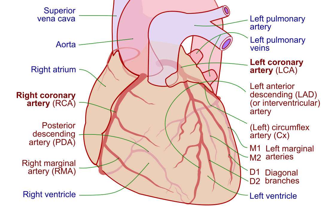
But coronary disease is only the beginning. Modern cath labs also measure pressures inside the heart and lungs to diagnose heart failure and valve disease, evaluate and treat heart block and slow rhythms by implanting a pacemaker, and — in electrophysiology labs — perform cardiac ablation to correct fast, abnormal rhythms such as atrial fibrillation, supraventricular tachycardia, and ventricular tachycardia. Some labs also close structural defects, repair or replace valves percutaneously, and support failing circulation with mechanical pumps.
- Diagnose coronary narrowing, valve gradients, filling pressures, and shunts.
- Treat blockages with angioplasty and stents, and reopen arteries fast in a heart attack.
- Manage rhythm by implanting pacemakers and defibrillators or ablating abnormal circuits.
- Support the circulation with temporary pumps when the heart is failing.
Inside the room: the equipment
The heart of the cath lab is a fluoroscopy system — an X-ray tube and flat-panel detector mounted on a movable C-shaped arm that swings around the patient to capture live, moving X-ray images from any angle. Everything else in the room is arranged around that imaging chain.
A patient table that glides in three directions sits at the center. Overhead and beside it hang banks of monitors showing the live X-ray, recorded angiographic runs, the ECG, and pressure waveforms. A shielded control room, separated by leaded glass, houses recording and monitoring workstations. On the sterile side, a small table holds the catheters, guidewires, sheaths, and the manifold — the cluster of stopcocks that lets one catheter switch between flushing, pressure monitoring, and contrast injection.
This overview touches only the essentials; the full inventory of sheaths, wires, catheters, imaging, and support devices is covered in our dedicated guide to the cath lab and its tools and the deeper equipment breakdown it links to. Here are the workhorses you will always find:
| Equipment | What it does |
|---|---|
| Fluoroscopy C-arm | Provides real-time X-ray imaging to guide catheters and film contrast injections |
| Patient table | Moves the patient precisely under the X-ray beam in three planes |
| Introducer sheath | Valved tube in the artery or vein that serves as the entry port for wires and catheters |
| Guidewires and catheters | Steer through vessels and engage the coronary arteries or heart chambers |
| Manifold and contrast injector | Deliver iodinated dye and switch between flush, pressure, and injection |
| Hemodynamic monitoring system | Displays and records ECG and pressure waveforms from every catheter |
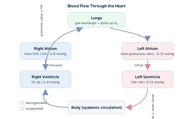
Those pressure waveforms are not decoration — they are the raw data of invasive hemodynamics. Learning to read them is a core skill for anyone working in the lab, and our hemodynamics study guide walks through what each tracing means, while the hemodynamic calculator handles the arithmetic behind measurements like cardiac output and resistance.
Who works in the cath lab: the team
A cath lab runs on a tightly coordinated team, and no single person could run a case alone. Each role has a defined job, and the choreography among them is part of what keeps a high-stakes procedure safe and fast.
At the sterile table stands the interventional cardiologist (or, for rhythm work, an electrophysiologist), who performs the catheterization — puncturing the vessel, steering the wires and catheters, and making the treatment decisions. Beside and around the physician, a supporting cast handles imaging, monitoring, medications, and documentation.
| Role | Primary responsibility |
|---|---|
| Interventional cardiologist / EP | Performs the catheterization, angiography, and intervention; leads clinical decisions |
| Cardiovascular technologist (RCIS) | Scrubs to assist at the table, monitors hemodynamics, operates recording systems, manages devices |
| Registered nurse | Administers medications and sedation, monitors the patient, tracks vital signs and comfort |
| Radiologic technologist | Operates the imaging equipment and helps optimize views while managing radiation dose |
| Circulating / control-room staff | Documents the case, hands supplies to the sterile field, runs the recording workstation |
The Registered Cardiovascular Invasive Specialist (RCIS) is the credential many technologists hold, and the role sits at the center of daily lab operations — reading pressure waveforms, recognizing rhythms on the monitor, preparing and passing devices, and anticipating the physician's next step. If you are exploring this path, our overview of the cardiovascular technologist career lays out the training and scope. Because the team must react instantly to the monitor, fluency in ECG interpretation and rhythm interpretation is non-negotiable — a sudden run of ventricular tachycardia or a dropped pressure changes the plan in seconds.
What happens before the procedure
Before a single catheter moves, the team completes a careful preparation sequence — because a smooth, safe case is set up long before the puncture. The patient is interviewed and examined, allergies (especially to iodinated contrast) are noted, and kidney function is checked because contrast is cleared by the kidneys.
Blood tests, an ECG, and often an echocardiogram are reviewed to build a picture of the heart before the operator ever looks inside. Blood thinners and diabetes medications may be adjusted, and the patient fasts for a period beforehand. Informed consent is obtained, with the risks and benefits explained plainly.
In the room, the patient lies on the table and is connected to continuous ECG, blood pressure, and oxygen monitoring. The access site — wrist or groin — is shaved if needed, cleaned with antiseptic, and draped in sterile fashion. Mild sedation is usually given so the patient is relaxed but awake enough to follow instructions like holding a breath. Finally, the team pauses for a time-out: confirming the patient, the procedure, the site, and any special concerns before starting. This safety checklist is standard of care and mirrors the surgical world.
- Screening — allergies, kidney function, current medications, and prior cardiac testing.
- Consent — risks, benefits, and alternatives discussed and documented.
- Preparation — monitoring leads placed, access site cleaned and draped, sedation started.
- Time-out — final team confirmation of patient, procedure, and site.
What happens during the procedure
The procedure itself follows a logical sequence: get access, steer to the heart, gather information, and — if needed — treat. Understanding this flow demystifies what can look like a blur of activity around the table.
Step one is vascular access. After numbing the skin with local anesthetic, the operator punctures the radial artery at the wrist (the modern default) or the femoral artery in the groin, then places a valved introducer sheath as the working port. Step two is navigation. A soft guidewire leads the way up the aorta, and a shaped catheter tracks over it until its tip seats in the opening of a coronary artery or crosses into a heart chamber.
Step three is diagnosis. Contrast dye is injected while the C-arm films the flow — this is the coronary angiogram, showing narrowings as they choke the dye column. Pressures are recorded from the catheter tip, and in selected lesions a pressure wire measures fractional flow reserve to judge whether a borderline narrowing is truly flow-limiting. Step four, when indicated, is intervention. A fine wire crosses the blockage, a balloon dilates it, and a drug-eluting stent is deployed to hold the artery open. Throughout, the patient may feel a warm flush from the dye or brief chest pressure during a balloon inflation, but the catheters themselves are not painful because the inside of blood vessels has no sensation.
Medications are woven through every step — anticoagulants to prevent clotting on the equipment, nitroglycerin to dilate vessels or relieve spasm, and antiplatelet drugs for stenting. Our reference on cath lab medications covers the common agents, and the specific role of nitroglycerin shows up constantly at the table. The whole diagnostic-versus-treatment decision tree is spelled out in PCI versus cardiac catheterization.
Types of procedures performed
Cath labs are not all the same — the term covers several kinds of labs that share the same X-ray backbone but do very different work. Knowing the categories helps you understand why one lab is quiet and methodical while another is a rapid-response heart-attack team.
| Procedure type | What it involves |
|---|---|
| Diagnostic coronary angiogram | Contrast imaging of the coronary arteries to find and grade narrowings |
| Percutaneous coronary intervention (PCI) | Balloon angioplasty and stenting to reopen a blocked coronary artery |
| Primary PCI for STEMI | Emergency artery reopening during an acute heart attack, done against the clock |
| Right-heart catheterization | Pressure and cardiac-output measurement from the right heart and lungs |
| Electrophysiology (EP) study and ablation | Mapping and destroying abnormal electrical circuits to fix arrhythmias |
| Device implantation | Placing pacemakers and defibrillators through the veins |
| Structural interventions | Percutaneous valve repair or replacement and defect closure |
The most time-critical is primary PCI for a STEMI — an ST-elevation myocardial infarction, where a coronary artery is completely blocked and heart muscle is dying by the minute. Recognizing that pattern on the tracing triggers the whole pathway, which is why reading a STEMI on the ECG is a genuine emergency skill; the underlying event is detailed in our guide to myocardial infarction. Right-heart catheterization, meanwhile, is the source of the pressures used to diagnose shock and cardiac tamponade and to calculate values through tools like the Fick cardiac output calculator.
Safety, radiation, and risks
Because the cath lab runs on X-rays and invades the vascular system, safety is engineered into everything — protecting both the patient and a team that spends every workday near a radiation source. Radiation protection follows three principles any RCIS candidate should be able to recite: time, distance, and shielding.
The team minimizes fluoroscopy time, keeps as much distance from the X-ray tube as possible (dose falls off with the square of distance), and uses shielding — lead aprons, thyroid collars, leaded eyewear, and movable barriers. Everyone wears a dosimeter to track cumulative exposure, and modern systems apply dose-reduction technology to keep both patient and operator dose as low as reasonably achievable.
Cardiac catheterization is generally very safe, but it is still an invasive procedure with real, if uncommon, risks: bleeding or bruising at the access site, allergic-type reactions to contrast, contrast-associated kidney injury, arrhythmias, and — rarely — vessel injury, stroke, or heart attack. Radial access has become the default partly because it lowers bleeding and access-site complications compared with the femoral approach. The full landscape of complications and how the team prevents and manages them is a subject in its own right, worth studying alongside the procedure.
- Time — image only when actively needed; store loops rather than taking extra runs.
- Distance — step back from the tube whenever possible; doubling distance quarters the dose.
- Shielding — lead apron, thyroid shield, leaded glasses, and movable barriers between staff and the source.
How a cath lab differs from surgery and other labs
A common point of confusion is how a cath lab relates to an operating room, and how a coronary cath lab differs from an electrophysiology lab. Clearing this up helps place the room in the broader map of cardiac care.
Unlike an operating room, a cath lab treats the heart from inside the blood vessels rather than by opening the chest — the whole appeal of a "percutaneous" (through-the-skin) approach is a small puncture, faster recovery, and no cardiac surgery for many conditions that once required it. That said, cath labs and cardiac surgery are partners, not rivals; complex disease is often discussed by a combined heart team, and hybrid rooms can do both.
Within the catheter world, the coronary/hemodynamic cath lab focuses on arteries, pressures, and structural work, while the electrophysiology (EP) lab focuses on the heart's wiring — mapping and ablating the circuits behind arrhythmias, and implanting rhythm devices. Both use catheters and fluoroscopy, but the EP lab adds specialized mapping systems and targets the cardiac conduction system rather than the coronary arteries. To ground either one, it helps to have the underlying cardiac anatomy and ECG fundamentals firmly in place, and to test yourself with focused hemodynamics practice questions.
Key takeaways
- A cardiac cath lab is an X-ray-guided procedure room where catheters are threaded through blood vessels to diagnose and treat heart disease without open surgery.
- It handles both diagnostic work (angiography and pressure measurement) and interventional work (balloon angioplasty and stenting), plus rhythm and structural procedures.
- The room centers on a fluoroscopy C-arm, a moving table, monitors for imaging and hemodynamics, and a sterile table of sheaths, wires, catheters, and the manifold.
- A coordinated team — interventional cardiologist, cardiovascular technologist (RCIS), nurse, and radiologic tech — each owns a defined role during the case.
- Procedures follow a four-step flow: access, navigate, diagnose, treat, with medications woven through every stage.
- Primary PCI for a STEMI is the most time-critical case; right-heart catheterization supplies the pressures behind shock and tamponade diagnoses.
- Safety rests on time, distance, and shielding for radiation, with radial access lowering bleeding risk and complications remaining uncommon but real.
- This content is educational, not medical advice, and cath lab practice evolves — always defer to current guidelines and the care team.
How long does a cath actually take?
See the full timeline from check-in to discharge.
See Cath Timing →Frequently asked questions
What is a cardiac cath lab?
A cardiac cath lab, or cardiac catheterization laboratory, is a specialized hospital procedure room built around a real-time X-ray imaging system. In it, physicians insert thin, flexible tubes called catheters into a blood vessel at the wrist or groin and steer them to the heart to diagnose and treat cardiovascular disease. It is used for coronary angiograms, balloon angioplasty and stenting, pressure measurements, rhythm procedures, and more — all through a small puncture rather than open surgery.
What is the difference between a cardiac catheterization and a cath lab?
The cath lab is the room and its equipment; cardiac catheterization is the procedure performed inside it. Catheterization means placing a catheter into a blood vessel and guiding it to the heart, whether to inject contrast for an angiogram, measure pressures, or deliver treatment. So a patient goes to the cath lab (the place) to have a cardiac catheterization (the procedure).
What equipment is in a cardiac cath lab?
Core equipment includes a fluoroscopy C-arm that provides live X-ray imaging, a patient table that moves in three planes, and banks of monitors showing the X-ray, ECG, and pressure waveforms. On the sterile side there are introducer sheaths, guidewires, and catheters, plus a manifold and contrast injector for dye. Interventional cases add balloons, drug-eluting stents, intracoronary imaging, and pressure wires, along with a hemodynamic monitoring and recording system.
Who works in a cardiac cath lab?
A cath lab team typically includes an interventional cardiologist (or electrophysiologist) who performs the procedure, a cardiovascular technologist — often credentialed as an RCIS — who assists at the table and monitors hemodynamics, a registered nurse who manages medications and patient monitoring, and a radiologic technologist who operates the imaging equipment. Control-room staff document the case and support the sterile field. Every case needs all these functions covered.
What happens during a cardiac catheterization?
The procedure follows four steps. First, the operator numbs the skin and places a sheath in the radial or femoral artery. Second, a guidewire and catheter are steered up to the heart under X-ray. Third, contrast dye is injected to film the coronary arteries and pressures are recorded — this is the diagnostic angiogram. Fourth, if a blockage needs treatment, a balloon dilates it and a stent is placed to hold the artery open. Patients are usually sedated but awake.
Is a cardiac catheterization painful?
Most patients feel little pain. The access site is numbed with local anesthetic, so the main sensation is brief pressure during the puncture. The catheters themselves are not painful because the inside of blood vessels has no pain sensation. Patients often feel a warm flush when contrast dye is injected, and may notice brief chest pressure during a balloon inflation, which passes quickly. Mild sedation keeps the patient relaxed throughout.
What is the difference between a cath lab and an EP lab?
Both use catheters and X-ray guidance, but they target different things. A coronary or hemodynamic cath lab focuses on the arteries, chamber pressures, and structural heart work — angiograms, stenting, and valve procedures. An electrophysiology (EP) lab focuses on the heart's electrical system, mapping and ablating the abnormal circuits behind arrhythmias and implanting pacemakers and defibrillators. Many hospitals have separate rooms for each, and some rooms can do both.
How long does a cardiac catheterization take?
A straightforward diagnostic coronary angiogram often takes roughly 20 to 45 minutes, though timing varies. If an intervention such as stenting is added, the case can run one to two hours or longer for complex disease. Patients then recover for a period afterward — usually shorter with radial (wrist) access than with femoral (groin) access, since the wrist site allows sitting up and walking sooner. Actual times depend on the patient and the procedure.
Is a cardiac cath lab procedure safe?
Cardiac catheterization is generally very safe and is done millions of times a year, but as an invasive, X-ray-guided procedure it carries small risks. These include bleeding or bruising at the access site, contrast allergy or contrast-associated kidney injury, arrhythmias, and rarely vessel injury, stroke, or heart attack. Radial access lowers bleeding complications, and the team uses radiation-protection principles of time, distance, and shielding. Serious complications are uncommon in experienced hands.
Sources & further reading
- Cardiovascular Credentialing International (CCI)
- American College of Cardiology
- American Heart Association
- MedlinePlus (U.S. National Library of Medicine)
External links are provided for reference; always confirm current details with the official source.