Shock Hemodynamics
Shock is not one disease but four distinct circulatory failures — and each writes its own signature across CVP, PCWP, cardiac output, and SVR. Learn to read that four-number fingerprint and you can name the type of shock at the bedside before a single lab result comes back.
- What shock actually is (and why hemodynamics defines it)
- The four numbers that decode every shock state
- The shock hemodynamics chart (CVP, PCWP, CO, SVR)
- Hypovolemic shock: the empty tank
- Cardiogenic shock: the failing pump
- Obstructive shock: flow blocked in the pipes
- Distributive shock: the pipes gone slack
- How these pressures are measured: the Swan-Ganz catheter
- Mixed shock and why patterns blur
- Shock hemodynamics on the RCIS exam
- Key takeaways
What shock actually is (and why hemodynamics defines it)
Shock is a state of circulatory failure in which oxygen delivery no longer meets the metabolic demand of the tissues. That mismatch — not a specific blood pressure number — is the true definition. A patient can be in shock with a "normal" systolic pressure if perfusion has quietly collapsed, and can look hypotensive without being in shock at all. What matters is whether cells are getting enough oxygen, which is why rising lactate, cool or mottled skin, falling urine output, and altered mentation are the clinical alarms that count.
Because oxygen delivery is the product of cardiac output and arterial oxygen content, almost every form of shock comes back to a failure of flow, a failure of the pump, an obstruction to filling or ejection, or a collapse of vascular tone. Those four failure modes map cleanly onto the four classic categories: hypovolemic, cardiogenic, obstructive, and distributive. The elegance of hemodynamic monitoring is that each category leaves a reproducible pattern in the pressures and flows we can measure.
This article is educational and written for RCIS candidates, cardiovascular technologists, and clinicians building their foundations — it is not medical advice, and real patients are managed by their own care teams using current institutional protocols. If you want the underlying physiology first, the RCIS hemodynamics guide lays out how pressures, flow, and resistance interlock before you layer shock on top.
The four numbers that decode every shock state
Four measurements do most of the diagnostic heavy lifting. Commit them to memory and you have a portable framework for any shock question on the RCIS exam or any confusing patient in the unit.
- CVP — central venous pressure, a surrogate for right atrial pressure and therefore right-heart preload. Normal is roughly 2–6 mmHg.
- PCWP — pulmonary capillary wedge pressure, the downstream estimate of left atrial pressure and left-ventricular preload. Normal wedge is about 6–12 mmHg.
- CO — cardiac output, the flow the pump generates, normally 4–8 L/min (indexed to 2.5–4.0 L/min/m² as the cardiac index).
- SVR — systemic vascular resistance, the afterload the left ventricle ejects against, normally 800–1200 dynes·sec·cm⁻⁵.
The trick is that shock changes these in predictable combinations. Lose volume and both filling pressures fall while resistance clamps up. Lose pump function and filling pressures back up while output drops. Lose vascular tone and resistance craters while output paradoxically rises. Once you see it as a pattern of four dials moving together, the memorization collapses into logic. Each of these values deserves its own study — the mechanics of cardiac output, the meaning of systemic vascular resistance, and how they scale to body size in the cardiac index.
The shock hemodynamics chart (CVP, PCWP, CO, SVR)
Here is the single most useful table in this whole topic. Print it, quiz yourself on it, and rebuild it from scratch until you can draw the arrows without looking. Nearly every RCIS shock question is a variation on reading one column of this chart.
| Shock type | CVP (preload) | PCWP (LV preload) | Cardiac output | SVR (afterload) |
|---|---|---|---|---|
| Hypovolemic | ↓ low | ↓ low | ↓ low | ↑ high |
| Cardiogenic | ↑ high | ↑ high | ↓ low | ↑ high |
| Obstructive (tamponade / PE) | ↑ high | variable* | ↓ low | ↑ high |
| Distributive (septic / warm) | ↓ low / normal | ↓ low / normal | ↑ high | ↓ low |
*In tamponade the diastolic pressures equalize, so CVP, PCWP, and pericardial pressure converge; in massive pulmonary embolism the wedge is often low-normal because the left heart is underfilled behind the obstruction.
Hypovolemic shock: the empty tank
Hypovolemic shock is a problem of volume. Hemorrhage, severe dehydration, burns, or third-spacing drains the intravascular compartment until there is simply not enough blood returning to fill the heart. With preload gone, both filling pressures fall — CVP low, PCWP low — and because stroke volume depends on adequate filling, cardiac output drops with them.
The compensatory response is dramatic and predictable. The sympathetic nervous system fires, arterioles constrict to defend blood pressure, and SVR climbs. Clinically this is the cold, pale, tachycardic patient with a thready pulse and delayed capillary refill. Early on, that vasoconstriction can hold blood pressure near normal even as substantial volume is lost, which is exactly why pressure alone is a poor early warning; the tachycardia and narrowing pulse pressure show up first. A quick bedside estimate of this compensation is the shock index — heart rate divided by systolic pressure — which rises before frank hypotension appears.
The physiologic fix is to refill the tank: restore intravascular volume with fluids or blood products, and control the source of loss. On monitoring, successful resuscitation shows filling pressures climbing back toward normal and cardiac output recovering. Current resuscitation guidance increasingly favors balanced strategies — measured fluid or blood rather than reflexive large-volume crystalloid — but the specifics evolve with the evidence, and any real protocol belongs to the treating team.
Cardiogenic shock: the failing pump
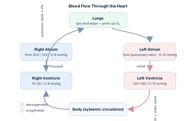
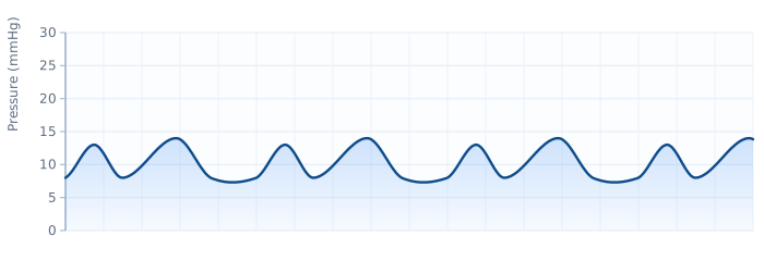
In cardiogenic shock the tank is full but the pump has failed. The commonest cause is a large myocardial infarction that knocks out enough left-ventricular muscle to cripple ejection; other causes include acute valve failure, end-stage cardiomyopathy, and malignant arrhythmias. Because the ventricle cannot empty, blood backs up behind it. Pressures rise upstream: PCWP climbs high, often above 18 mmHg, and as the right heart congests too, CVP rises. Meanwhile forward flow collapses, so cardiac output falls and the body clamps down, driving SVR up.
The classic bedside picture is "cold and wet": cold from the low output and high resistance, wet from the pulmonary congestion the high wedge produces. Rising wedge pressure is the reason these patients drown in their own lungs — fluid is pushed across the pulmonary capillaries into the alveoli. Recognizing the underlying infarct quickly is everything, which is why fluency in STEMI ECG interpretation and the broader picture of myocardial infarction is inseparable from managing this shock.
Therapeutically the goals run opposite to hypovolemia: unload the congested ventricle and support forward flow rather than pour in fluid. That may mean inotropes, careful diuresis, and mechanical support such as the intra-aortic balloon pump, which lowers afterload and boosts coronary perfusion. Emergency revascularization for the culprit infarct remains the intervention with the strongest survival benefit, and where and how it is done is covered in PCI versus cardiac catheterization.
Obstructive shock: flow blocked in the pipes
Obstructive shock is a mechanical problem: the heart and blood volume may be fine, but something physically blocks filling or ejection. The three headline causes are cardiac tamponade, tension pneumothorax, and massive pulmonary embolism, and understanding each requires knowing where the block sits.
In cardiac tamponade, fluid in the pericardial sac squeezes the heart from outside. The chambers cannot fill, so diastolic pressures rise and — crucially — equalize: CVP, right-ventricular diastolic pressure, PA diastolic, and PCWP all converge toward the same elevated number. Output falls, resistance rises, and pulsus paradoxus appears. The pattern is distinctive enough that cardiac tamponade hemodynamics is worth studying as its own case; the fix is drainage, not fluid alone.
In massive pulmonary embolism, a clot obstructs the pulmonary arteries. The right ventricle strains against a suddenly enormous afterload, CVP rises as the right heart backs up, but the left heart is underfilled behind the block, so the wedge is typically low-normal — a useful clue that distinguishes PE from tamponade. Cardiac output falls and SVR rises. The common thread across obstructive shock is high CVP with low output; the treatment is always to relieve the obstruction — pericardiocentesis, needle decompression, or clot lysis — because fluids and pressors alone cannot fix a mechanical dam.
Distributive shock: the pipes gone slack
Distributive shock is the odd one out, and the RCIS exam loves to test it precisely because its numbers run backward from the others. The problem is not volume or pump but tone: the vasculature dilates massively, resistance collapses, and blood pools in an overlarge vascular space. Sepsis is the dominant cause; anaphylaxis, neurogenic shock from spinal injury, and adrenal crisis round out the group.
Because the vessels are wide open, SVR falls dramatically — the single most defining feature. To compensate, the heart often speeds up and pumps harder, so early distributive shock frequently shows a high cardiac output. Filling pressures (CVP and PCWP) are usually low or normal, partly from the enlarged vascular space and partly from capillary leak pulling fluid out of the circulation. This produces the classic "warm shock": flushed, warm extremities with bounding pulses, in stark contrast to the cold, clamped skin of the other three types.
Management centers on restoring tone and treating the cause: source control and antibiotics for sepsis, vasopressors such as norepinephrine to reconstrict the vasculature, and judicious fluid to fill the expanded space. Because vasopressor and vasodilator choices are a recurring theme in the cath lab, the cath-lab medications overview is a natural companion to this section.
How these pressures are measured: the Swan-Ganz catheter
The numbers in the shock chart are not abstractions — they come from real measurements, most classically the pulmonary artery catheter. The Swan-Ganz (pulmonary artery) catheter is floated from a central vein through the right atrium and right ventricle into the pulmonary artery. Along the way it reads CVP in the right atrium, right-ventricular and pulmonary artery pressures, and then, when the balloon is wedged in a distal pulmonary artery, the pulmonary capillary wedge pressure that estimates left atrial filling.
Cardiac output is obtained by thermodilution through the same catheter, or calculated by the Fick principle from oxygen consumption. SVR is never measured directly; it is calculated from mean arterial pressure, CVP, and cardiac output. If you want to see how those inputs combine, our hemodynamic calculator lets you plug in pressures and watch resistance and index fall out, and the Fick cardiac output calculator handles the oxygen-based route.
Routine pulmonary artery catheterization has fallen out of favor for many patients — large trials failed to show a survival benefit from reflexive PA-catheter use, and less invasive monitoring has grown up alongside it. But in complex or mixed shock the catheter still earns its keep by revealing exactly which of the four dials is deranged. Reading its tracings is a core cath-lab skill; the pressure-waveform work in the hemodynamics guide and hands-on hemodynamics practice questions are the fastest way to build that fluency.
Mixed shock and why patterns blur
Real patients rarely read the textbook. A septic patient who has also been bleeding, or an infarct patient who develops sepsis from a line infection, produces a mixed hemodynamic picture where the clean arrows of the chart pull against each other. A septic patient with septic cardiomyopathy may show the low SVR of distribution alongside a depressed output that mimics cardiogenic shock. This is precisely where invasive monitoring changes decisions: it tells you whether to give fluid, squeeze the vessels, support the pump, or relieve an obstruction — actions that would harm the wrong shock type.
Two habits keep you out of trouble. First, always ask what the SVR and cardiac output are doing together, because that pair separates the vasodilated states from the low-flow states faster than any single number. Second, treat the chart as a starting hypothesis, not a verdict — re-measure after each intervention and watch the dials move. A wedge that keeps climbing after fluids, or a resistance that will not rise on pressors, is telling you the diagnosis is shifting.
It also helps to keep the neighboring concepts sharp. Falling output can stem from a failing stroke volume or a collapsing ejection fraction, and arrhythmias that trigger or worsen shock — from ventricular tachycardia to fast atrial fibrillation — are diagnosed on the same monitors, so ECG interpretation and shock reading go hand in hand.
Shock hemodynamics on the RCIS exam
Registry questions on shock are remarkably formulaic once you know the chart. The classic item hands you a set of values — a CVP, a wedge, an output, and an SVR — and asks you to name the shock type. Work it in a fixed order and you will rarely miss: (1) look at cardiac output and SVR first, because low-output/high-SVR versus high-output/low-SVR splits the field in half; (2) then use the filling pressures to separate within each half — low filling pressures point to hypovolemia, high filling pressures point to cardiogenic or obstructive; (3) finally, use the clue words (equalized diastolic pressures for tamponade, warm skin for sepsis) to lock it in.
Round out your preparation with the flow and resistance fundamentals, then drill the numbers under timed conditions. The RCIS hemodynamics question bank mixes shock patterns with waveform reading, and pairing it with the hemodynamics guide gives you both the why and the how. Because shock so often rides on top of an arrhythmia or an infarct, spending time on ECG practice questions pays off on exam day too.
Key takeaways
- Shock is failed oxygen delivery, not a specific blood pressure — lactate, mentation, urine output, and skin tell the real story.
- Four numbers decode it: CVP (right preload), PCWP (left preload), cardiac output (flow), and SVR (afterload).
- Hypovolemic: everything down (CVP, PCWP, CO) except a high, compensating SVR — refill the tank.
- Cardiogenic: high filling pressures (CVP and PCWP up), low output, high SVR — "cold and wet"; unload and revascularize.
- Obstructive: high CVP with low output; tamponade equalizes diastolic pressures, PE spares the wedge — relieve the block.
- Distributive: the rebel — low SVR with high or normal output, "warm shock" — restore tone and treat the source.
- Read output and SVR together first, then use filling pressures and clue words to pinpoint the type.
- This is educational material for exam prep and clinical foundations, not medical advice; real management follows current guidelines and the treating team.
Practise hemodynamics
Test shock, pressures, and waveform questions with explanations.
Practise Hemodynamics →Frequently asked questions
What are the four types of shock and their hemodynamics?
The four categories are hypovolemic, cardiogenic, obstructive, and distributive. Hypovolemic shows low CVP, low PCWP, low cardiac output, and high SVR. Cardiogenic shows high CVP, high PCWP, low output, and high SVR. Obstructive shows high CVP, low output, and high SVR (with equalized diastolic pressures in tamponade and a low-normal wedge in pulmonary embolism). Distributive shows low or normal CVP and PCWP, high or normal cardiac output, and a distinctively low SVR.
What is the shock hemodynamics chart?
It is a quick-reference table showing how CVP, PCWP, cardiac output, and SVR move in each type of shock. It lets you name the shock type from four numbers: three of the four (hypovolemic, cardiogenic, obstructive) share a low output and high SVR, while distributive shock stands apart with high output and low SVR.
How do CVP and PCWP differ?
CVP (central venous pressure) reflects right atrial pressure and right-heart preload, normally about 2–6 mmHg. PCWP (pulmonary capillary wedge pressure) is measured by wedging a balloon-tipped catheter in the pulmonary artery and estimates left atrial pressure and left-ventricular preload, normally about 6–12 mmHg. Together they report filling on both sides of the heart.
Why is SVR high in most types of shock but low in distributive shock?
In hypovolemic, cardiogenic, and obstructive shock the body senses falling perfusion and constricts arterioles to defend blood pressure, driving SVR up. Distributive shock is caused by loss of vascular tone itself — sepsis, anaphylaxis, or neurogenic injury dilate the vessels — so SVR falls, which is the defining fingerprint of this category.
How do you tell cardiogenic from hypovolemic shock hemodynamically?
Look at the filling pressures. Both show low cardiac output and high SVR, but hypovolemic shock has low CVP and low PCWP (an empty tank), while cardiogenic shock has high CVP and high PCWP because blood backs up behind a failing pump. High wedge means cardiogenic; low wedge means hypovolemic.
What is 'warm shock'?
Warm shock is the early distributive (usually septic) picture: because the vasculature is dilated and SVR is low while cardiac output is high, the skin is warm and flushed with bounding pulses, unlike the cold, clamped extremities of the other shock types. Late septic shock can convert to a cold, low-output state as the heart is depressed.
How is cardiac tamponade different from other obstructive shock?
Tamponade compresses all four chambers equally from outside, so the diastolic pressures — CVP, right and left ventricular diastolic, PA diastolic, and PCWP — rise and equalize toward the same value, often with pulsus paradoxus. Massive pulmonary embolism, by contrast, raises CVP but leaves the wedge low-normal because the left heart is underfilled behind the clot. The treatment for tamponade is pericardial drainage, not fluid alone.
Do you still need a Swan-Ganz catheter to diagnose shock?
Not routinely. Large trials failed to show a survival benefit from reflexive pulmonary artery catheter use, and less invasive monitoring plus echocardiography now cover many cases. The catheter still earns its place in complex or mixed shock, where directly reading CVP, PA pressures, wedge, and thermodilution output reveals exactly which parameter is deranged.
What is the first step when reading shock hemodynamics on the RCIS exam?
Look at cardiac output and SVR together before anything else. Low output with high SVR points to hypovolemic, cardiogenic, or obstructive shock, while high or normal output with low SVR points to distributive shock. Once you have split the field that way, use the filling pressures and clue words to pinpoint the exact type.
Sources & further reading
- Cardiovascular Credentialing International (CCI)
- American College of Cardiology
- American Heart Association
- MedlinePlus (U.S. National Library of Medicine)
External links are provided for reference; always confirm current details with the official source.