Intra-Aortic Balloon Pump (IABP): How It Works, Timing & Waveform
The intra-aortic balloon pump (IABP) is the workhorse of temporary mechanical circulatory support — a balloon that inflates and deflates in perfect rhythm with the heart to unload the left ventricle and drive blood into the coronary arteries. This guide breaks down counterpulsation physiology, timing on the arterial waveform, indications, complications, and the exam-ready details every cardiovascular technologist needs.
What is an intra-aortic balloon pump?
An intra-aortic balloon pump is a catheter-mounted, elongated balloon positioned in the descending thoracic aorta that inflates during diastole and deflates during systole to support a failing left ventricle. It is the oldest and still the most widely used form of temporary mechanical circulatory support, first used clinically in the late 1960s and refined into the reliable bedside device seen in cardiac cath labs, cardiac ICUs, and cardiac surgery suites today.
The balloon itself is made of polyethylene and holds roughly 30 to 50 mL of helium, sized to the patient. It rides on a slim catheter that is usually inserted through the femoral artery and advanced retrograde until the tip sits just below the origin of the left subclavian artery, with the balloon body spanning the descending aorta above the renal arteries. Helium is used as the shuttle gas because it is light, moves quickly in and out of the balloon, and is harmlessly absorbed if the balloon ever ruptures.
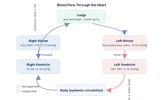
What makes the device clever is that it adds no pumping force of its own. It has no impeller and moves no blood directly. Instead, it borrows the patient's own cardiac cycle and simply times a volume of gas to help at exactly the right moments. That principle is called counterpulsation, and it is the heart of everything that follows. If you want the pressure-and-flow foundations behind it, our hemodynamics study guide lays the groundwork.
This article is educational and not medical advice. Device settings, indications, and thresholds vary by patient and institution; always follow current guidelines and local protocols.
How counterpulsation works
Counterpulsation means the balloon does the opposite of the ventricle: it inflates when the heart relaxes and deflates when the heart contracts. Two mechanical events happen on every beat, and together they explain the entire clinical benefit.
Diastolic augmentation. The instant the aortic valve closes and the ventricle begins to relax, the balloon inflates. Because the aorta is a closed, fluid-filled space, displacing 40 mL of volume upward pushes a bolus of blood back toward the aortic root and the coronary ostia. This raises aortic pressure during diastole — the phase when the coronary arteries actually fill — and improves myocardial oxygen supply. On the arterial trace this shows up as a tall spike right after the dicrotic notch.
Systolic unloading (afterload reduction). Just before the ventricle contracts, the balloon deflates rapidly. Emptying 40 mL creates a sudden low-pressure sink in the aorta, so the ventricle ejects against markedly less resistance. Lower afterload means the heart does less work, uses less oxygen, and empties more completely, which nudges stroke volume and cardiac output upward.
The elegance is that the IABP improves the oxygen-supply side and the oxygen-demand side at once. Coronary flow goes up while cardiac workload goes down — precisely the combination a struggling, ischemic ventricle needs. Understanding the shape of the aortic pressure pulse it manipulates makes the timing rules that follow far more intuitive.
| Cardiac phase | Balloon action | Hemodynamic effect |
|---|---|---|
| Diastole (ventricle relaxing) | Inflates | Raises diastolic aortic pressure → more coronary perfusion (supply ↑) |
| Systole (ventricle contracting) | Deflates | Lowers aortic pressure just before ejection → afterload and workload ↓ (demand ↓) |
The physiologic benefits, quantified
It helps to see what the device actually buys the patient. The magnitude of each effect depends on balloon volume, heart rate, aortic compliance, and how well the timing is dialed in, but the direction of every change is consistent.
- Coronary perfusion pressure rises because diastolic aortic pressure is augmented, delivering more oxygen to the myocardium — the main reason the IABP relieves ongoing ischemia.
- Left-ventricular afterload falls, reducing wall stress and myocardial oxygen consumption. The ventricle ejects against a lower pressure, so it works less hard for the same output.
- Cardiac output improves modestly, typically on the order of 0.5 to 1 L/min. That is meaningful support, but it is a boost — not a full replacement for a failing pump.
- Left-ventricular filling pressures ease, which can lower pulmonary capillary wedge pressure and relieve pulmonary congestion.
Two limits are worth stating plainly. First, the IABP depends on a heart that is still ejecting; it augments a rhythm, it does not create one. In a nearly motionless or fibrillating ventricle, its benefit shrinks. Second, the cardiac-output gain is smaller than what newer trans-valvular flow pumps deliver, which is one reason patient selection has narrowed over the past decade. You can review how these numbers fit into overall pump performance in our discussions of cardiac output and cardiac index.
Reading the IABP timing waveform
Timing is where the IABP is won or lost, and the arterial pressure waveform is the tool that gets it right. When the pump is set to a 1:2 assist ratio — augmenting every other beat — the monitor displays assisted and unassisted beats side by side, and comparing them reveals whether inflation and deflation are landing correctly.
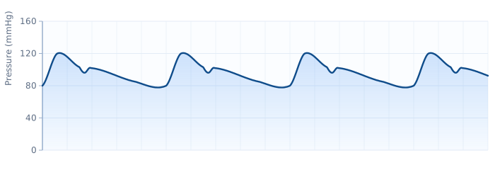
The dicrotic notch is the reference point. That small notch on the downslope of the arterial trace marks aortic valve closure and the start of diastole — the moment inflation should begin. Correct inflation produces a sharp V shape at the notch and a diastolic augmentation peak that is higher than the patient's own unassisted systolic pressure. Deflation should occur just before the next systole, dropping the end-diastolic pressure and lowering the following assisted systolic peak compared with the unassisted one.
On a well-timed trace you can point to four favorable findings: augmented diastolic pressure exceeding unassisted systole, a reduced balloon-aortic end-diastolic pressure, a lower assisted systolic pressure, and a clean inflation right at the dicrotic notch. Miss any of these and the timing needs adjustment.
| Timing error | What you see on the waveform | Consequence |
|---|---|---|
| Early inflation | Balloon inflates before the dicrotic notch; augmentation encroaches on systole | Aortic valve loaded prematurely, ↑ afterload, ↓ stroke volume, potential aortic regurgitation |
| Late inflation | Inflation appears well after the notch; a distinct notch is still visible before augmentation | Suboptimal diastolic augmentation, reduced coronary perfusion benefit |
| Early deflation | Sharp U-shaped dip, then pressure rises again before systole | Poor afterload reduction, possible retrograde coronary flow and angina |
| Late deflation | Balloon-aortic end-diastolic pressure equal to or above unassisted end-diastole; widened augmentation | Balloon obstructs ejection, dramatically ↑ afterload and myocardial demand — the most dangerous error |
Practicing waveform recognition on a monitor is a core RCIS skill, and it dovetails with the arterial-tracing work in our hemodynamics guide. Many candidates find it helps to pair this with rhythm reading, since the pump can be triggered off the ECG as well.
Triggering and assist ratios
The console has to know when each cardiac cycle begins so it can time the balloon. It picks up that cue from a trigger, and the choice of trigger matters at the bedside.
- ECG trigger. The most common mode. The R wave marks systole, and the console times deflation to the R wave and inflation to the middle of the T wave. Clean electrode contact is essential, which is why familiarity with the ECG and lead placement pays off.
- Arterial pressure trigger. The console uses the upstroke of the arterial waveform to identify systole. Useful when the ECG signal is poor, but it needs an adequate pulse pressure — typically a systolic of at least 50 mmHg.
- Pacer and internal triggers. Special modes handle paced rhythms or, as a fallback, run at a fixed internal rate when no reliable physiologic trigger exists.
Assist ratio describes how many beats the balloon augments. A 1:1 ratio augments every beat and gives maximal support; 1:2 augments every other beat and is the standard setting for weaning and for waveform timing checks; 1:3 provides minimal support and is used late in the weaning process. Weaning is done by stepping the ratio down and watching whether the patient's own hemodynamics hold — never by simply switching the pump off, since a stationary balloon in the aorta is a thrombosis risk.
Arrhythmias challenge triggering. An irregular rhythm such as atrial fibrillation makes beat-to-beat timing unpredictable, and very fast rates like supraventricular tachycardia shorten diastole so much that augmentation becomes inefficient. Modern consoles include algorithms that adapt to irregular rhythms, but recognizing the rhythm remains a clinician's job — a skill our ECG rhythm interpretation guide is built to sharpen.
Indications: when the IABP is used
The IABP is a bridge, not a destination. It supports the circulation while a reversible problem is fixed or while a longer-term plan is arranged. The established indications cluster around ischemia, pump failure, and mechanical complications.
- Cardiogenic shock, classically after a large myocardial infarction, when the ventricle cannot maintain adequate output despite drugs.
- Refractory unstable angina and ongoing ischemia not controlled by medical therapy, where diastolic augmentation buys coronary perfusion.
- Mechanical complications of MI, such as acute mitral regurgitation from papillary-muscle rupture or a ventricular septal defect, where afterload reduction stabilizes the patient before surgery.
- High-risk or complicated percutaneous coronary intervention, as hemodynamic backup during a difficult procedure — the intersection of catheter and pump discussed in our overview of PCI versus cardiac catheterization.
- Bridge to surgery, transplant, or a durable device, and support for weaning from cardiopulmonary bypass in the operating room.
Two evidence points deserve honesty. The IABP-SHOCK II trial did not show a mortality benefit from routine IABP use in cardiogenic shock complicating MI, and as a result guidelines no longer recommend it reflexively for that setting. It remains valuable for mechanical complications and as a bridge, and it is still chosen for individual patients based on judgment. This is an area where the evidence is genuinely evolving, and newer support devices are reshaping practice. The broader picture of shock hemodynamics is worth reviewing alongside any decision to place one.
Contraindications and cautions
Some patients should not receive an IABP, and knowing the absolute limits is a favorite exam theme because they follow directly from how the device works.
| Absolute contraindications | Why |
|---|---|
| Moderate-to-severe aortic regurgitation | Diastolic augmentation drives blood backward through the incompetent valve, worsening regurgitation and ventricular loading |
| Aortic dissection | Balloon inflation in a dissected aorta can extend the tear or rupture the vessel |
| Aortic aneurysm (large) | Risk of rupture and embolization of mural thrombus |
| Severe peripheral arterial disease | May prevent safe insertion and threatens limb perfusion; a graft or end-stage vessel can be uncrossable |
Relative cautions include severe uncontrolled coagulopathy or bleeding, uncontrolled sepsis, small-caliber or heavily calcified iliofemoral vessels, and severe tachyarrhythmias that undermine reliable triggering. The single most important pattern to memorize is that aortic regurgitation is an absolute contraindication — because the balloon's diastolic push is exactly the wrong thing for a leaky aortic valve. If you are shaky on why, revisiting the mechanics in the counterpulsation section above makes it click.
Complications to watch for
Because the catheter sits in a large artery and inflates a balloon dozens of times a minute, the complications are predominantly vascular, hematologic, and mechanical. Vigilance is a nursing and technologist priority throughout the support period.
- Limb ischemia is the most common serious complication. The femoral catheter can obstruct distal flow, so pulses, color, temperature, and sensation in the cannulated leg are checked frequently. A cold, pulseless, painful limb demands immediate action.
- Bleeding and hematoma at the insertion site, made worse by the anticoagulation often used to prevent catheter thrombus.
- Thrombocytopenia, because the balloon mechanically shears platelets with every cycle; platelet counts are trended.
- Balloon rupture, signaled by blood in the helium line or a loss of augmentation. It requires urgent removal because helium embolism and, more dangerously, entrapment from clotted blood can occur.
- Malposition, a balloon that migrates too high can occlude the left subclavian or carotid vessels; too low, it can obstruct the renal arteries and drop urine output. Position is confirmed on chest imaging.
- Aortic injury, dissection, infection, and stroke round out the less common but serious risks.
A sudden loss of the diastolic augmentation waveform is always worth a fast, structured check: is it a trigger problem, a timing problem, a kink, low balloon volume, or a rupture? Reading that waveform is the fastest window into whether the device — and the patient — is in trouble.
IABP versus other support devices
The IABP no longer stands alone. A newer generation of percutaneous devices provides more flow, and knowing where the balloon pump sits in that landscape is increasingly tested and clinically relevant.
| Device | Mechanism | Flow support | Notes |
|---|---|---|---|
| IABP | Counterpulsation in the aorta | ~0.5–1 L/min boost | Easiest to place, lowest cost, needs native ejection; no mortality benefit shown in MI shock |
| Trans-valvular axial-flow pump | Continuous flow from LV to aorta | Up to 3.5–5.5 L/min | More powerful, larger sheath, higher hemolysis and vascular risk |
| Percutaneous VA-ECMO | External oxygenator and pump | Full cardiopulmonary support | Supports lungs and heart; most invasive, resource-intensive |
The practical distinction: the IABP assists a beating heart, while the flow pumps and ECMO can substitute for one. That is why the balloon remains a first-line choice for ischemia, mechanical MI complications, and moderate support needs, while profound shock increasingly moves to higher-output options. The right choice is patient-specific and institution-specific, and the field is shifting quickly enough that guidelines are revised often.
For technologists building the bigger picture, it helps to connect the IABP to the environment it lives in — the cardiac cath lab — and to the invasive monitoring that runs alongside it, such as the Swan-Ganz catheter used to track filling pressures and cardiac output during support. Solidify the surrounding anatomy with our cardiac anatomy guide, then test yourself on the hemodynamics practice questions.
Key takeaways
- The intra-aortic balloon pump is a helium-filled balloon in the descending aorta that provides temporary circulatory support through counterpulsation.
- It inflates in diastole to augment coronary perfusion (supply ↑) and deflates in systole to reduce afterload and myocardial workload (demand ↓) — improving supply and demand at once.
- The dicrotic notch is the timing landmark; correct inflation sits right at the notch and augmented diastolic pressure should exceed unassisted systole.
- The four timing errors — early/late inflation and early/late deflation — each have a recognizable waveform, and late deflation is the most dangerous because the balloon obstructs ejection.
- Core indications include cardiogenic shock, refractory ischemia, mechanical MI complications, high-risk PCI, and bridging; routine use in MI shock is no longer guideline-recommended after IABP-SHOCK II.
- Aortic regurgitation and aortic dissection are absolute contraindications, flowing directly from how diastolic augmentation works.
- Limb ischemia is the most common serious complication; watch the cannulated leg, platelet count, waveform, and balloon position closely.
- This content is educational, not medical advice, and mechanical-support practice is evolving — always defer to current guidelines and local protocols.
Practise hemodynamics & devices
Test IABP, waveforms, and pressure questions with explanations.
Practise Hemodynamics →Frequently asked questions
What does an intra-aortic balloon pump do?
An IABP supports a failing left ventricle through counterpulsation. It inflates in the aorta during diastole to push blood back toward the coronary arteries, increasing myocardial oxygen supply, and deflates during systole to lower the pressure the ventricle ejects against, reducing cardiac workload. It improves oxygen supply and lowers oxygen demand at the same time, typically adding about 0.5 to 1 L/min of cardiac output.
When does the balloon inflate and deflate?
The balloon inflates during diastole, timed to the dicrotic notch when the aortic valve closes and the coronary arteries fill. It deflates during systole, just before the ventricle contracts, to create a low-pressure sink that eases ejection. A helpful memory aid is that inflation belongs to diastole and deflation belongs to systole.
Why is helium used in an IABP?
Helium is used because it is a very light, low-density gas that moves in and out of the balloon rapidly, allowing fast inflation and deflation to keep pace with the heart. It is also inert and highly soluble, so if the balloon ruptures the gas is absorbed and cleared with far less risk of a dangerous embolism than a heavier gas would pose.
What is the dicrotic notch and why does it matter for IABP timing?
The dicrotic notch is the small downstroke on the arterial pressure waveform that marks aortic valve closure and the start of diastole. It is the timing landmark for the IABP: correct inflation occurs right at the notch, producing a sharp V shape and a diastolic augmentation peak that rises above the patient's unassisted systolic pressure. Inflating before or after the notch causes the classic timing errors.
What are the four IABP timing errors?
The four errors are early inflation, late inflation, early deflation, and late deflation. Early inflation loads the aortic valve prematurely and raises afterload; late inflation gives poor diastolic augmentation; early deflation causes a dip-and-rebound that wastes afterload reduction; and late deflation is the most dangerous because the balloon is still inflated as the ventricle tries to eject, sharply increasing afterload and myocardial demand.
What are the main indications for an IABP?
The IABP is used for cardiogenic shock, refractory unstable angina and ongoing ischemia, mechanical complications of myocardial infarction such as acute mitral regurgitation or a ventricular septal defect, hemodynamic support during high-risk PCI, and as a bridge to surgery, transplant, or a durable device. It also helps wean patients from cardiopulmonary bypass.
What are the contraindications to an intra-aortic balloon pump?
Absolute contraindications include moderate-to-severe aortic regurgitation, aortic dissection, and a large aortic aneurysm, because diastolic augmentation would worsen these conditions. Severe peripheral arterial disease is also a contraindication when it prevents safe insertion or threatens limb perfusion. Relative cautions include uncontrolled coagulopathy, sepsis, and severe tachyarrhythmias that disrupt triggering.
What is the most common complication of an IABP?
Limb ischemia in the cannulated leg is the most common serious complication, because the femoral catheter can obstruct distal blood flow. Clinicians frequently check the pulse, color, temperature, and sensation of the affected limb. Other important complications include bleeding at the insertion site, thrombocytopenia from platelet shearing, balloon rupture, and malposition that can block the subclavian or renal arteries.
Does an IABP improve survival in cardiogenic shock?
The evidence is nuanced. The IABP-SHOCK II trial found no mortality benefit from routine IABP use in cardiogenic shock complicating myocardial infarction, so guidelines no longer recommend it reflexively in that setting. It remains valuable for mechanical complications of MI and as a bridge, and it is still chosen for selected patients. This is an evolving area, and newer higher-output support devices are increasingly used for profound shock.
Sources & further reading
- Cardiovascular Credentialing International (CCI)
- American College of Cardiology
- American Heart Association
- MedlinePlus (U.S. National Library of Medicine)
External links are provided for reference; always confirm current details with the official source.