Cardiac Cath Lab Equipment: The Essential Tools
Walk into any cardiac cath lab and you are surrounded by a purpose-built toolkit — sheaths, guidewires, catheters, a manifold, contrast injectors, and intracoronary imaging and physiology systems — each engineered for one job in a tightly choreographed procedure. This guide breaks down the essential cardiac cath lab equipment every cardiovascular technologist needs to know, from the femoral sheath to IVUS, OCT, FFR, and the intra-aortic balloon pump.
- The cath lab environment: what the equipment has to work with
- Vascular access and sheaths
- Guidewires: steering through the vasculature
- Diagnostic and guiding catheters
- The manifold, contrast, and injection system
- Balloons, stents, and interventional devices
- Intracoronary imaging: IVUS and OCT
- Coronary physiology: FFR and iFR
- Hemodynamic monitoring and support devices
- Imaging hardware and radiation safety
- Key takeaways
The cath lab environment: what the equipment has to work with
Cardiac cath lab equipment is the coordinated set of catheters, wires, sheaths, imaging, and monitoring devices used to access the vascular system, navigate to the heart, and diagnose or treat cardiovascular disease. Before diving into individual tools, it helps to picture the room they live in. A modern cath lab centers on a fluoroscopy C-arm that shoots real-time X-ray images, a patient table that moves in three planes, a bank of monitors displaying live imaging and hemodynamic data, and a control room shielded from radiation.
Everything in the sterile field is designed to be introduced through a small puncture in an artery or vein, threaded through the vasculature under fluoroscopic guidance, and manipulated with millimeter precision. That constraint — long, thin, steerable, and radiographically visible — shapes the design of every catheter and wire in the room. If you want the full tour of the space itself, our overview of what a cardiac cath lab is sets the scene, and the distinction between diagnostic and interventional work is covered in PCI versus cardiac catheterization.
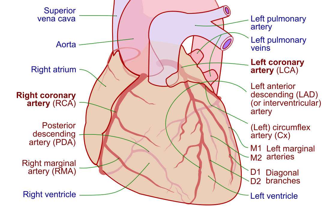
A quick note before we start naming devices: mastering the equipment is inseparable from knowing the coronary artery anatomy it navigates and the pressures it measures. The tools only make sense against that anatomical and physiologic backdrop.
This article is educational and not medical advice. Device choices, sizes, and techniques vary by patient and institution; always follow current guidelines and local protocols.
Vascular access and sheaths
The introducer sheath is the gateway to the entire procedure — a short, valved tube placed in the artery or vein that lets catheters and wires pass in and out without bleeding or losing access. Nothing else happens until a sheath is in.
Access begins with a percutaneous puncture, classically using the Seldinger technique: a needle enters the vessel, a guidewire is threaded through the needle, the needle is removed, and the sheath is advanced over the wire. The two dominant arterial access sites are the radial artery at the wrist and the femoral artery in the groin. Radial access has become the default for most diagnostic and interventional coronary work in current practice because it lowers bleeding and vascular complications and lets patients sit up sooner; femoral access remains important for larger-bore devices, complex interventions, and mechanical support.
Sheaths are sized by their inner diameter in French units, where 3 French equals roughly 1 millimeter. A 6 French sheath — inner diameter of about 2 mm — handles most diagnostic and routine PCI work. Larger 7 to 8 French or bigger sheaths are used when a bulky device such as an atherectomy system, a large-bore imaging catheter, or a circulatory-support pump must pass through. The French size stamped on the sheath refers to the largest catheter it will accept.
| Access site | Typical use | Trade-offs |
|---|---|---|
| Radial (wrist) | Default for diagnostic and most PCI | Fewer bleeding complications, faster ambulation; smaller vessel limits very large devices, risk of radial spasm or occlusion |
| Femoral (groin) | Large-bore devices, complex PCI, mechanical support | Accommodates big sheaths; higher bleeding and access-site complication risk, longer bed rest |
Guidewires: steering through the vasculature
Guidewires are the rails the catheters ride on — thin, steerable wires that lead the way through arteries, around bends, and across lesions so that catheters can follow. A catheter almost never travels alone; it tracks over a wire that has already found the path.
Cath labs stock a spectrum of wires for different jobs. A 0.035-inch J-tip guidewire is the standard workhorse for advancing diagnostic catheters up the aorta; its floppy, curved tip is atraumatic and slides past plaque without gouging the vessel wall. For coronary intervention, much finer 0.014-inch coronary guidewires are used — soft-tipped, torqueable wires that a cardiologist can steer into a specific branch and push across a blockage. Specialized wires add stiffness for crossing hard chronic total occlusions, extra support for delivering stents, or hydrophilic coatings for slippery tortuous anatomy.
Three properties define how a wire behaves: tip stiffness (how much force the tip delivers, which trades crossing power against perforation risk), body support (how well the wire's shaft backs up device delivery), and coating (hydrophilic for slickness or hydrophobic for tactile feedback). Choosing among them is a craft, but the underlying goal is always the same: reach the target safely and give following devices a stable track.
- 0.035" J-tip — advancing diagnostic catheters through the aorta; soft J shape avoids vessel trauma.
- 0.014" workhorse coronary wire — routine PCI, balancing steerability and support.
- Stiff / CTO wires — penetrating hard, calcified, or totally occluded segments.
- Extra-support wires — anchoring device delivery in difficult anatomy.
Diagnostic and guiding catheters
Catheters are the shaped, hollow tubes that engage the coronary ostia, deliver contrast, measure pressure, and act as conduits for interventional devices. Their preformed curves are the reason a cardiologist can seat a catheter tip precisely into the left or right coronary artery from a wrist or groin puncture.
It helps to split them into two families. Diagnostic catheters are used for angiography and pressure measurement; they have thinner walls and shapes optimized to selectively engage a vessel and inject dye. The classic examples are the Judkins Left and Judkins Right curves for the left and right coronary arteries, plus pigtail catheters for ventriculography and aortic root shots. Guiding catheters, used in intervention, have a larger lumen and stiffer walls so they can deliver balloons, stents, and other hardware while providing the back-up support needed to push devices across a lesion.
| Catheter | Primary use | Notes |
|---|---|---|
| Judkins Left (JL) | Engaging the left main coronary artery | Sized by the curve length, e.g. JL4; most common left diagnostic shape |
| Judkins Right (JR) | Engaging the right coronary artery | JR4 is the everyday right-sided choice |
| Pigtail | Left ventriculography, aortography | Coiled tip distributes contrast and reduces jet injury during power injection |
| Guiding catheter | Delivering PCI devices | Larger lumen, stiffer body for support; e.g. EBU, XB, Amplatz shapes |
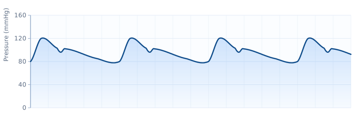
Diagnostic catheters also carry pressure. When the tip sits in the aorta, left ventricle, or a great vessel, it transmits a pressure waveform back to the transducer — the raw material of invasive hemodynamics. That link between catheter position and waveform is central to the hemodynamics study guide, and the pressures a pigtail records in the left ventricle and aorta underpin measurements like the aortic valve area.
The manifold, contrast, and injection system
The manifold is the control hub at the sterile table — a set of stopcocks and tubing that lets the operator switch a single catheter between pressure monitoring, contrast injection, saline flush, and blood aspiration without ever disconnecting. It is the unglamorous piece of equipment that ties the whole hemodynamic and imaging workflow together.
A typical manifold has three or more ports connected by stopcocks: one line runs to the pressurized flush (heparinized saline) to keep the catheter patent, one to the transducer for continuous pressure display, and one to the contrast reservoir for hand injections. By turning stopcocks, the operator selects which function reaches the catheter tip. Careful manifold technique is also how air bubbles are kept out of the system — a small but serious safety discipline, since an air embolus injected into a coronary artery can cause ischemia.
Iodinated contrast media is what makes the coronary tree and chambers visible under X-ray. Modern labs use low- or iso-osmolar nonionic agents to reduce discomfort and adverse reactions. Contrast can be delivered by hand through the manifold or by a power injector for high-volume, high-pressure shots such as ventriculography and aortography. Because contrast carries risks — allergic-type reactions and contrast-associated kidney injury among them — dose is tracked and minimized, especially in patients with reduced renal function. The properties, risks, and mitigation strategies are detailed in our dedicated piece on iodinated contrast media.
Balloons, stents, and interventional devices
Once a lesion is crossed with a wire, the interventional hardware comes into play. These are the tools that actually open a narrowed artery in percutaneous coronary intervention, treating the coronary artery disease that brought the patient to the lab.
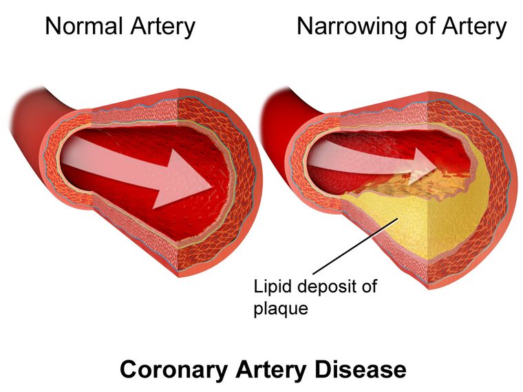
- Angioplasty balloons mount on the coronary wire and inflate at the lesion to compress plaque and dilate the vessel. They come as compliant, semi-compliant, and non-compliant types, and specialty versions add cutting or scoring edges for resistant plaque.
- Drug-eluting stents (DES) are the mainstay of modern PCI — a metal scaffold coated with an antiproliferative drug that props the artery open and releases medication to prevent re-narrowing (restenosis). They have largely displaced bare-metal stents.
- Atherectomy devices — rotational, orbital, or laser — sand down or ablate heavily calcified plaque so a balloon or stent can be delivered.
- Intracoronary lithotripsy uses sonic pressure waves to crack calcium, a newer tool for stubborn calcified lesions where the evidence base is still growing.
- Thrombectomy (aspiration) catheters remove clot, used selectively rather than routinely under current guidance.
These devices are why timely PCI is the treatment of choice for an acute myocardial infarction, where a blocked artery must be reopened fast. Reading the ECG that triggers that pathway — recognizing a STEMI on the ECG — is the upstream skill that sends a patient to this equipment in the first place.
Intracoronary imaging: IVUS and OCT
Intravascular ultrasound (IVUS) and optical coherence tomography (OCT) are catheter-based imaging tools that look inside the artery wall — something angiography, which only shows the lumen silhouette, cannot do. They have moved from research curiosities to everyday tools for optimizing complex PCI.
Both use a miniature imaging element on a catheter that is pulled back through the segment of interest to build a cross-sectional picture of the vessel. The difference is the energy source. IVUS uses sound waves: it penetrates deeper into the vessel wall, sees through blood without flushing, and is excellent for sizing the vessel, assessing plaque burden, and guiding stent expansion, especially in the left main and larger vessels. OCT uses near-infrared light: it offers far higher resolution — fine enough to see stent struts, dissections, and thin plaque caps — but penetrates less deeply and requires a brief contrast flush to clear blood from the field.
| Feature | IVUS (ultrasound) | OCT (light) |
|---|---|---|
| Energy source | Sound waves | Near-infrared light |
| Resolution | Lower (about 100–150 microns) | Very high (about 10–20 microns) |
| Penetration depth | Deeper — sees full vessel wall | Shallower |
| Blood clearing | Not required | Requires contrast/saline flush |
| Best strengths | Vessel sizing, plaque burden, left main, renal-sparing | Stent apposition, edge dissection, fine detail |
Current guidelines increasingly endorse intravascular imaging to guide PCI in complex and high-risk lesions, where randomized data suggest imaging-guided stenting improves outcomes over angiography alone. The choice between IVUS and OCT is often a matter of the lesion, the vessel, and operator preference — and in patients with impaired kidneys, IVUS has the advantage of needing no extra contrast. This is an evolving area, and adoption continues to expand as the evidence matures.
Coronary physiology: FFR and iFR
Fractional flow reserve (FFR) is a pressure-wire measurement that tells you whether a given narrowing is actually starving the muscle of blood — turning a picture of a blockage into a functional verdict. Angiography shows how tight a lesion looks; FFR shows whether it matters.
The technique uses a coronary guidewire with a tiny pressure sensor near the tip. The operator measures pressure distal to the lesion and compares it with pressure in the aorta. FFR is calculated during maximal hyperemia — vessels dilated fully, usually with intravenous or intracoronary adenosine — as the ratio of distal coronary pressure to aortic pressure. A value at or below 0.80 generally indicates a hemodynamically significant lesion that may benefit from stenting; values above that suggest the narrowing can be managed medically. Deferring PCI on a lesion that is not flow-limiting spares the patient an unnecessary stent.
A newer family of resting indices, most notably the instantaneous wave-free ratio (iFR), measures a pressure ratio during a specific quiet window of diastole without needing adenosine, which avoids the drug's side effects and speeds the assessment. Large trials have shown resting indices to be non-inferior to FFR for guiding revascularization decisions, and both approaches are endorsed in current practice. Because these measurements are built on the same distal-and-aortic pressure logic used throughout invasive hemodynamics, they slot naturally alongside the pressure concepts in our hemodynamics guide and the arithmetic in the hemodynamic calculator.
Hemodynamic monitoring and support devices
Beyond diagnosis and intervention, the cath lab houses equipment for measuring pressures and, when the heart falters, for supporting the circulation. A hemodynamic monitoring system ties every fluid-filled catheter to a transducer and displays live pressure waveforms — the same signals that let you calculate cardiac output, derive systemic vascular resistance, and interpret filling pressures at the bedside.
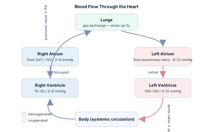
The classic right-heart tool is the balloon-tipped, flow-directed pulmonary artery catheter. Advanced through a central vein, it records right atrial, right ventricular, pulmonary artery, and pulmonary capillary wedge pressures, and it measures cardiac output by thermodilution. Its role, waveforms, and pitfalls are covered in depth in our guide to the Swan-Ganz catheter, and the diagnostic patterns it reveals are central to reading shock hemodynamics and cardiac tamponade.
When the ventricle cannot keep up, the lab reaches for temporary mechanical circulatory support. The oldest and most common is the intra-aortic balloon pump (IABP), a helium-filled balloon in the descending aorta that inflates in diastole to boost coronary perfusion and deflates in systole to reduce afterload — the counterpulsation physiology explained fully in our intra-aortic balloon pump article. Higher-output percutaneous flow pumps and veno-arterial ECMO provide more support for profound shock. Device selection is patient-specific and the evidence around routine use continues to shift, so guidelines are revised often.
Imaging hardware and radiation safety
None of the catheters or wires would be usable without the imaging chain that makes them visible, and that same X-ray comes with a dose the whole team must respect. The fluoroscopy system — an X-ray tube and flat-panel detector on a C-arm — provides the real-time imaging that guides every movement, while cine angiography captures high-quality recorded runs of the coronary injections for review and measurement.
Radiation protection rests on three principles that any RCIS candidate should be able to recite: time, distance, and shielding. Minimize fluoroscopy time, maximize distance from the source (intensity falls off with the square of distance), and use shielding — lead aprons, thyroid collars, leaded glasses, and table-mounted and ceiling-suspended barriers. Staff wear dosimeters to track cumulative exposure, and modern systems use dose-reduction technology and framing techniques to keep patient and operator dose as low as reasonably achievable.
- Time — keep the fluoro pedal down only when actively imaging; store fluoro loops instead of taking extra cine runs when possible.
- Distance — step back when you can; the inverse-square law means doubling your distance quarters the dose.
- Shielding — lead apron, thyroid shield, leaded eyewear, and movable barriers between you and the tube.
Handling this equipment competently is a defining part of the cardiovascular technologist's role. If you are mapping out the career and credential, our overview of the cardiovascular ultrasound technologist pathway and the broader RCIS material give the bigger picture.
Key takeaways
- Vascular access starts with a valved introducer sheath, sized in French units (French ÷ 3 ≈ mm), placed via radial (default) or femoral (large-bore) access.
- Guidewires lead and catheters follow — a 0.035" J-tip for diagnostic navigation and 0.014" steerable wires for coronary intervention, chosen by tip stiffness, support, and coating.
- Catheters split into diagnostic shapes (Judkins Left/Right, pigtail) that engage vessels and transmit pressure, and larger-lumen guiding catheters that deliver PCI hardware.
- The manifold switches a catheter between flush, pressure monitoring, and contrast injection; aspirate-then-flush discipline keeps air and thrombus out of the coronaries.
- IVUS (sound) penetrates deeper and needs no flush; OCT (light) gives higher resolution but needs a contrast flush — both refine complex PCI beyond what angiography shows.
- FFR ≤ 0.80 (measured at maximal hyperemia) flags a flow-limiting lesion worth treating; resting indices like iFR reach a similar decision without adenosine.
- The IABP and higher-output pumps provide temporary circulatory support, while pulmonary artery catheters and transducers supply the hemodynamic data.
- Respect radiation with time, distance, and shielding. This content is educational, not medical advice, and cath lab practice evolves — always defer to current guidelines and local protocols.
Practise cath-lab fundamentals
Test equipment, procedures, and device questions with explanations.
Practise RCIS Core →Frequently asked questions
What equipment is used in a cardiac cath lab?
Core cardiac cath lab equipment includes vascular access sheaths, guidewires, diagnostic and guiding catheters, a manifold with stopcocks for flushing and injecting, iodinated contrast and a power injector, and a fluoroscopy C-arm with monitors. Interventional cases add angioplasty balloons and drug-eluting stents, intracoronary imaging (IVUS and OCT), pressure wires for FFR, and hemodynamic monitoring and support devices such as the intra-aortic balloon pump.
What is the difference between a sheath, a guidewire, and a catheter?
A sheath is the short valved tube placed in the artery or vein that serves as the entry port for everything else. A guidewire is a thin, steerable wire that finds the path through the vessels and across lesions. A catheter is the hollow, preshaped tube that tracks over the guidewire to engage a vessel, inject contrast, measure pressure, or deliver interventional devices. In short: the sheath is the doorway, the wire leads, and the catheter follows.
What is a manifold in the cath lab?
A manifold is a set of stopcocks and tubing at the sterile table that connects a single catheter to several lines at once — a pressurized saline flush, a pressure transducer, and a contrast reservoir. By turning the stopcocks, the operator switches the catheter among flushing, pressure monitoring, and contrast injection without disconnecting anything. Careful manifold technique, including aspirating before injecting, keeps air bubbles and clot out of the coronary arteries.
What is the difference between IVUS and OCT?
Both are catheter-based intracoronary imaging tools, but they use different energy. IVUS uses ultrasound (sound waves), penetrates deeper into the vessel wall, and works without clearing blood, making it strong for vessel sizing and assessing plaque burden. OCT uses near-infrared light, giving much higher resolution to see stent struts and dissections in fine detail, but it penetrates less deeply and needs a brief contrast flush to clear blood from the field.
What does FFR measure and what is a normal value?
Fractional flow reserve (FFR) uses a pressure-sensing guidewire to measure whether a coronary narrowing is actually limiting blood flow. It is the ratio of pressure beyond the lesion to pressure in the aorta, measured during maximal hyperemia induced with a drug such as adenosine. An FFR value at or below 0.80 generally indicates a flow-limiting lesion that may benefit from stenting, while higher values suggest the narrowing can be treated medically.
What is the difference between FFR and iFR?
FFR is measured during maximal hyperemia, meaning a vasodilator like adenosine must be given to fully dilate the vessels before the pressure ratio is taken, with a treatment threshold around 0.80. The instantaneous wave-free ratio (iFR) is a resting index measured during a specific quiet phase of diastole without any hyperemic drug, which avoids adenosine's side effects and speeds the assessment. Large trials show resting indices are non-inferior to FFR for guiding revascularization.
Why is radial access preferred over femoral access?
Radial access at the wrist has become the default for most diagnostic and interventional coronary procedures because it produces fewer bleeding and access-site complications and lets patients sit up and walk sooner. Femoral access in the groin is still important when a large-bore device is needed — such as certain atherectomy systems, big imaging catheters, or circulatory support pumps — because the femoral artery accommodates larger sheaths than the smaller radial artery.
What is an introducer sheath and how is French size measured?
An introducer sheath is a short tube with a one-way valve placed in the vessel so catheters and wires can be exchanged without bleeding or losing access. Sheaths are sized in French units based on the largest catheter they accept, where 3 French is roughly 1 millimeter, so dividing the French number by 3 estimates the diameter in millimeters. A 6 French sheath handles most routine work, while larger devices require 7 French or bigger.
How does the cath lab protect against radiation exposure?
Radiation protection follows three principles: time, distance, and shielding. Staff minimize fluoroscopy time, keep as much distance from the X-ray source as possible (dose falls off with the square of distance), and use shielding such as lead aprons, thyroid collars, leaded eyewear, and movable barriers. Team members also wear dosimeters to track cumulative exposure, and modern systems apply dose-reduction technology to keep patient and operator exposure as low as reasonably achievable.
Sources & further reading
- Cardiovascular Credentialing International (CCI)
- American College of Cardiology
- American Heart Association
- MedlinePlus (U.S. National Library of Medicine)
External links are provided for reference; always confirm current details with the official source.