Cardiovascular Ultrasound Technologist
A cardiovascular ultrasound technologist is the allied-health specialist who captures moving pictures of the beating heart with sound waves — the person behind nearly every echocardiogram a cardiologist reads.
- What is a cardiovascular ultrasound technologist?
- Echocardiographer, cardiac sonographer, or ultrasound technologist?
- What does a cardiac sonographer do all day?
- Credentials: RCS, RDCS, and how to earn them
- Cardiac ultrasound vs invasive cardiology
- Cardiovascular ultrasound technologist salary and outlook
- The skills and knowledge that set great sonographers apart
- How to become a cardiac sonographer
- Key takeaways
What is a cardiovascular ultrasound technologist?
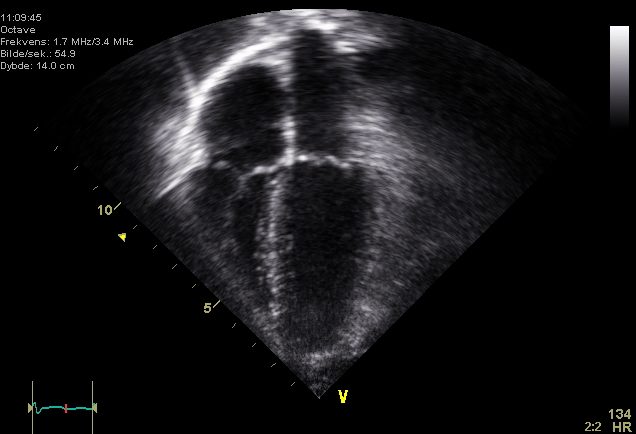
A cardiovascular ultrasound technologist is a trained imaging professional who uses high-frequency sound waves to picture the heart and, in some roles, the blood vessels. When the focus is specifically the heart, the same person is usually called an echocardiographer or a cardiac sonographer — the titles are effectively interchangeable in everyday practice. If you have ever had a technician slide a warm probe across your ribs while a gray, fan-shaped image of your heart pulsed on a screen, you have met one.
The work sits at the meeting point of physics, anatomy, and bedside care. A sonographer positions the patient, chooses the right acoustic window between the ribs, angles the transducer to open up each cardiac view, and optimizes the settings so a cardiologist can measure chamber size, wall motion, valve behavior, and blood-flow velocities. It is quiet, meticulous, radiation-free work that carries real diagnostic weight — a poorly acquired study can hide disease, and a well-acquired one can change a treatment plan the same afternoon.
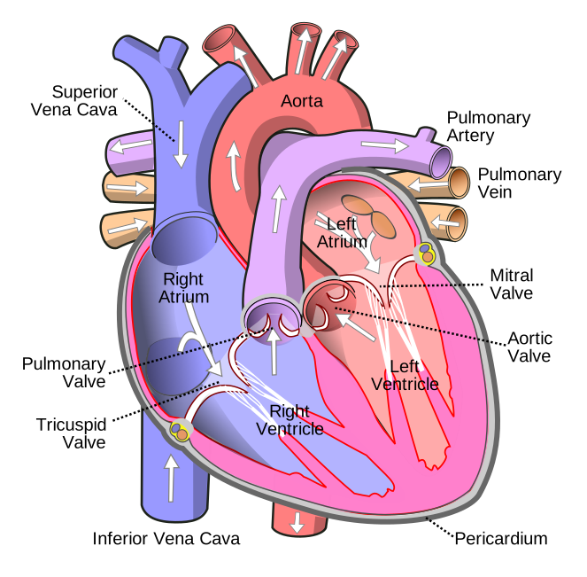
This role belongs to the broader family of the cardiovascular technologist, an umbrella that also covers invasive cath-lab and vascular specialists. Understanding cardiac anatomy in three dimensions is the foundation of the job, because a sonographer is essentially reconstructing a solid, moving organ from thin ultrasound slices in real time.
This article is educational, written for students, career-changers, and clinicians preparing for exams such as the RCIS and the sonography registries. It is not medical or career-placement advice; program requirements, scope, and pay vary by region, employer, and year.
Echocardiographer, cardiac sonographer, or ultrasound technologist?
Newcomers often trip over the overlapping job titles, so it helps to untangle them. All three describe someone who images the heart with ultrasound, but the emphasis differs slightly.
- Cardiac sonographer — the most common everyday term in the United States. It emphasizes the ultrasound (sonography) skill set and is the title most job postings use.
- Echocardiographer — literally "one who records the heart with sound." Some hospitals reserve this word for physicians who interpret echoes, so context matters. In many settings, though, it is used for the technologist who performs the study.
- Cardiovascular ultrasound technologist — a broader, more formal label. It signals that the person may image both the heart and peripheral vasculature, and it maps neatly onto the way credentialing and education programs describe the field.
In practice, the daily task is the same: acquire diagnostic-quality images and Doppler data of the heart. The differences live mostly in scope, employer preference, and which credential a person holds. That is where the alphabet soup of registries comes in.
What does a cardiac sonographer do all day?
A typical day is a string of scheduled studies punctuated by urgent bedside requests. The core task never changes — produce a complete, interpretable echocardiogram — but the settings range from a calm outpatient lab to a chaotic intensive-care unit. For the anatomy behind every view, a good echocardiogram primer is worth revisiting often.
Concretely, a cardiac sonographer will:
- Prepare and position the patient, explain the exam, and place ECG leads so each image is timed to the cardiac cycle.
- Acquire the standard windows — parasternal long- and short-axis, apical four- and two-chamber, and subcostal views — angling the probe to open each plane cleanly.
- Run the full toolkit of modes: 2D for structure, M-mode for precise line measurements, color Doppler to see leaks and shunts, and spectral (pulsed- and continuous-wave) Doppler to quantify velocities and estimate pressures.
- Measure on the fly — chamber dimensions, wall thickness, valve gradients, and the numbers that feed calculations such as ejection fraction, stroke volume, and aortic valve area.
- Recognize what is abnormal, capture extra clips of anything that looks off, and flag critical findings — a large pericardial effusion, a new severe valve leak, or a plunging pump function — to the reading physician right away.
Beyond the standard transthoracic study, sonographers often assist with transesophageal echocardiography (TEE), stress echoes tied to a cardiac stress test, contrast studies, and increasingly with strain imaging and 3D reconstruction. The best technologists are part physicist, part anatomist, and part detective.
Credentials: RCS, RDCS, and how to earn them
Two credentials dominate cardiac sonography in the United States, and both signal that a technologist has proven their knowledge and hands-on skill through a national exam.
| Credential | Issuing body | Full name | Focus |
|---|---|---|---|
| RDCS | ARDMS (American Registry for Diagnostic Medical Sonography) | Registered Diagnostic Cardiac Sonographer | Adult echo (AE) or pediatric echo (PE) specialty exams |
| RCS | CCI (Cardiovascular Credentialing International) | Registered Cardiac Sonographer | Adult echocardiography |
| RCCS | CCI | Registered Congenital Cardiac Sonographer | Pediatric / congenital echo |
Both the RCS and the RDCS are widely respected, and many employers accept either. Candidates typically qualify through one of two paths: completing an accredited cardiovascular or sonography program, or accumulating documented clinical experience and then sitting the registry exams. Most exams pair a general physical-principles component (ultrasound physics and instrumentation) with a heart-specific specialty component.
Because the field values demonstrated competence, credentialing is more than a formality. It affects hiring, pay, and the range of studies you are allowed to perform. It also builds directly on the same cardiac fundamentals that credentialing bodies test elsewhere — the conduction system, valve mechanics, and the pressure relationships covered in any solid hemodynamics guide.
Cardiac ultrasound vs invasive cardiology
One of the clearest ways to understand this career is to contrast it with its invasive cousin. A cardiac sonographer works with sound; an invasive cardiovascular technologist works inside the body, threading catheters through arteries in the cath lab. The vs invasive comparison shapes almost everything about the two roles — the risk, the environment, and the personality that thrives in each.
| Feature | Cardiac ultrasound (non-invasive) | Invasive cardiology |
|---|---|---|
| Primary tool | Ultrasound transducer | Catheters, guidewires, X-ray fluoroscopy |
| Radiation | None | Ionizing radiation (lead protection required) |
| Invasiveness | Non-invasive (skin contact only) | Invasive (arterial/venous access) |
| Typical setting | Echo lab, ICU, bedside, clinic | Cardiac catheterization lab |
| Data produced | Structure, function, flow velocities, estimated pressures | Directly measured pressures, cardiac output, angiograms |
| Pace | Deliberate, image-quality focused | Fast, procedural, sometimes emergent |
The two disciplines are complementary, not competing. An echo can estimate a valve gradient non-invasively; a catheter can measure the pressure directly. When findings disagree or a procedure is planned, patients move between both worlds. Anyone curious about the invasive side can explore what a cardiac cath lab is and how a diagnostic catheterization compares with PCI. The directly measured numbers from that side — right-heart pressures, the wedge, and calculated cardiac output — are exactly the values echo tries to approximate from the outside.
Cardiovascular ultrasound technologist salary and outlook
Compensation is one of the most searched questions about this career, and for good reason — cardiac sonography pays well relative to the training time. In the United States, cardiac sonographers and cardiovascular technologists generally earn a median in the broad range of roughly $65,000 to $95,000 per year, with experienced, credentialed sonographers in high-cost regions or specialized labs earning more. Travel and per-diem sonographers can command notably higher hourly rates.
Several factors move the number:
- Credentials — holding an RCS or RDCS (or both) tends to lift pay and open more positions.
- Geography — metropolitan and high-cost-of-living areas pay more, though the premium often tracks local expenses.
- Setting — hospitals, especially those with busy TEE and structural-heart programs, often pay above outpatient clinics.
- Experience and subspecialty — pediatric/congenital echo, TEE, and 3D/strain expertise are worth a premium.
The outlook is favorable. Employment for cardiovascular technologists and sonographers is projected to grow faster than the average for all occupations over the coming decade, driven by an aging population and the steadily expanding role of echocardiography in diagnosis and structural-heart procedures. Because these figures shift year to year, treat any single number as a snapshot; a deeper look at the cardiovascular technologist salary and the field's job outlook will give a fuller, more current picture.
The skills and knowledge that set great sonographers apart
Acquiring a diagnostic echo is harder than it looks. The heart hides behind ribs and lungs, it never stops moving, and every patient's anatomy is different. The technologists who stand out combine manual dexterity with deep cardiac understanding.
- Ultrasound physics — knowing why gain, depth, focus, and frequency change the image, and how Doppler angle affects velocity accuracy. This is half of every registry exam.
- Three-dimensional anatomy — mentally rotating the heart to predict where a structure will appear as you tilt the probe.
- Hemodynamic literacy — understanding how pressures and flows relate, so a measured velocity translates into a meaningful gradient. The same logic underlies systemic vascular resistance and cardiac index.
- ECG and rhythm recognition — because images are timed to the cardiac cycle and arrhythmias distort measurements. A sonographer who can read an ECG catches problems others miss; brushing up on ECG fundamentals pays off directly.
- Pattern recognition of disease — spotting a wall segment that barely moves after a heart attack, the flapping leaflet of severe regurgitation, or the swinging heart of a large effusion.
Rhythm awareness deserves emphasis. If a patient is in atrial fibrillation during the study, beat-to-beat variability forces the sonographer to average measurements across several cycles. Recognizing something ominous — a run of ventricular tachycardia or the pattern of a STEMI on the monitor — can turn a routine scan into a rapid-response call. This blend of imaging skill and clinical vigilance is what elevates a technologist from competent to indispensable.
How to become a cardiac sonographer
There is no single road, but the common routes share the same milestones: education, clinical training, and credentialing. Most people follow one of these paths.
- Accredited program — an associate or bachelor's degree, or a certificate for those already holding a health-science degree, in cardiovascular technology or diagnostic medical sonography with a cardiac concentration. This is the cleanest route and usually includes supervised clinical hours.
- Clinical-experience path — some candidates enter from a related allied-health role, accumulate documented scanning experience under supervision, and then qualify to sit the registry exams.
Whichever path you take, the finish line is the same: passing the physics component and a heart-specific specialty exam to earn the RCS, RDCS, or both. Programs weight cardiac anatomy, ultrasound physics, and pathology heavily, and success on the boards leans on the same core science tested across cardiology credentials — so working through practice questions in hemodynamics and ECG interpretation builds transferable strength. For a step-by-step overview of the broader field, the guides on how to become a cardiovascular technologist and the required schooling map out the timeline in detail.
A practical tip: shadow a working sonographer before committing. The job rewards patience, physical stamina (scanning is ergonomically demanding), and a genuine interest in the heart's mechanics. If those appeal to you, few allied-health careers offer as direct a line from training to meaningful, well-compensated work.
Key takeaways
- A cardiovascular ultrasound technologist — also called an echocardiographer or cardiac sonographer — images the beating heart with sound waves, producing the echocardiograms cardiologists rely on.
- The leading credentials are the RCS (from CCI) and the RDCS (from ARDMS); both prove competence and influence pay and scope.
- Cardiac ultrasound is non-invasive and radiation-free, in contrast to invasive cath-lab work that uses catheters and X-ray fluoroscopy — the two are complementary sides of cardiology.
- US salary typically falls in the roughly $65,000–$95,000 range, rising with credentials, subspecialty, and region; job growth outpaces the average occupation.
- Standout sonographers pair manual skill with ultrasound physics, 3D anatomy, hemodynamic reasoning, and rhythm recognition.
- The path runs through an accredited program or documented clinical experience, ending in the physics and cardiac specialty registry exams.
- This overview is educational and not medical or career-placement advice; verify current pay, scope, and program requirements locally.
Frequently asked questions
Is a cardiovascular ultrasound technologist the same as an echocardiographer?
Effectively yes. "Cardiovascular ultrasound technologist," "echocardiographer," and "cardiac sonographer" all describe someone who images the heart with ultrasound. The label "cardiovascular ultrasound technologist" is a bit broader and can include vascular imaging, while "cardiac sonographer" is the most common everyday US term. Note that some hospitals use "echocardiographer" for the interpreting physician, so confirm the meaning in context.
What is the difference between RCS and RDCS?
Both are cardiac sonography credentials earned by passing a national exam. The RDCS (Registered Diagnostic Cardiac Sonographer) is issued by ARDMS and has adult and pediatric echo specialties. The RCS (Registered Cardiac Sonographer) is issued by Cardiovascular Credentialing International (CCI). Many employers accept either, and some sonographers hold both to broaden their options.
Does a cardiac sonographer use radiation?
No. Cardiac ultrasound uses high-frequency sound waves, not ionizing radiation, so there is no radiation exposure and no lead apron required. That is a key contrast with invasive cardiovascular technologists in the cath lab, who work under X-ray fluoroscopy and must wear lead protection and dosimeters.
How much does a cardiovascular ultrasound technologist make?
In the United States, cardiac sonographers and cardiovascular technologists generally earn a median in the broad range of about $65,000 to $95,000 per year. Credentials such as the RCS or RDCS, subspecialties like TEE or pediatric echo, hospital settings, and high-cost regions push earnings toward the top of that range. Published figures are broad and change yearly, so treat any single number as a snapshot.
How long does it take to become a cardiac sonographer?
Most people finish in about one to two years through an accredited program — a shorter certificate if they already hold a health-science degree, or an associate degree otherwise — followed by the registry exams. Bachelor's routes and clinical-experience paths can take longer. The exact timeline depends on the program format and how quickly you complete required clinical hours.
Is echocardiography invasive?
Standard transthoracic echocardiography is non-invasive — the probe only touches the skin. Some specialized studies are semi-invasive, such as transesophageal echocardiography (TEE), where a probe is passed down the esophagus under sedation to image the heart from behind. Even TEE is far less invasive than catheter-based procedures in the cath lab.
What skills do you need to be a cardiac sonographer?
You need a strong grasp of ultrasound physics and instrumentation, three-dimensional cardiac anatomy, and basic hemodynamics, plus the manual dexterity to angle the probe into clear acoustic windows. ECG and rhythm recognition matter because images are timed to the cardiac cycle, and pattern recognition helps you catch abnormal findings. Patience, physical stamina, and good patient communication round out the profile.
Do cardiac sonographers diagnose heart disease?
Sonographers acquire and measure the images, flag critical or abnormal findings, and often recognize disease patterns, but the formal diagnosis and interpretation are made by a physician (typically a cardiologist) who reads the study. A skilled sonographer's judgment during acquisition strongly shapes the quality of that final interpretation.
Can a cardiac sonographer move into the cath lab or vascular imaging?
Yes. The cardiovascular technology field is broad, and many professionals cross-train. A cardiac sonographer might add vascular ultrasound credentials or, with additional training, move toward invasive roles in the catheterization lab. Because all these paths share a foundation in cardiac anatomy, hemodynamics, and ECG, skills transfer well between them.
Sources & further reading
- Cardiovascular Credentialing International (CCI)
- American College of Cardiology
- American Heart Association
- MedlinePlus (U.S. National Library of Medicine)
External links are provided for reference; always confirm current details with the official source.