Echocardiogram: What It Shows, Types & What to Expect
An echocardiogram is an ultrasound scan of the heart — a painless test that shows the heart's structure and function in real time. Here's what it reveals, the difference between an echo and an ECG, and what happens during the test.
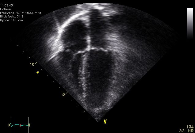
What is an echocardiogram?
An echocardiogram (echo) is an ultrasound scan that uses sound waves to create moving images of the heart. It is non-invasive and radiation-free, and it shows the heart beating in real time — the chambers, the valves, and the flow of blood through them. It is one of the most useful tests in cardiology.
What an echocardiogram shows
- Ejection fraction and overall pumping (systolic) function — see our ejection fraction guide
- Chamber size and wall thickness (hypertrophy or dilation)
- Valve function — stenosis or regurgitation
- Wall motion abnormalities from ischaemia or infarction
- Pericardial effusion and signs of tamponade
- Blood flow and pressures using Doppler
TTE vs TEE
| Transthoracic (TTE) | Transesophageal (TEE) | |
|---|---|---|
| Probe | On the chest wall | Down the esophagus (behind the heart) |
| Comfort | Painless, no prep | Sedation; throat numbing |
| Best for | General assessment | Valves, left atrium, clots, endocarditis |
A stress echo adds imaging during exercise or with dobutamine to unmask ischaemia.
Echocardiogram vs ECG
They sound alike but test completely different things:
| Echocardiogram | ECG (EKG) | |
|---|---|---|
| Measures | Structure & function | Electrical activity |
| Technology | Ultrasound images | Electrodes / tracing |
| Shows | Valves, EF, wall motion | Rhythm, rate, ischaemia |
Learn the electrical side in our ECG interpretation guide. The two tests are complementary, not interchangeable.
How long it takes & what to expect
A standard transthoracic echo takes about 30–60 minutes. You lie on your side while a cardiac sonographer moves a gel-covered probe across your chest. It's painless, needs no preparation, and there's no radiation. A cardiologist then interprets the images.
Key takeaways
- An echocardiogram is a painless ultrasound of the heart.
- It shows ejection fraction, chamber size, valves, and wall motion.
- TTE is done on the chest; TEE goes down the esophagus for clearer valve/atrial views.
- An echo images structure; an ECG records electrical activity — different tests.
- A standard echo takes about 30–60 minutes.
Interested in performing echoes?
See the cardiovascular ultrasound technologist career path.
Explore the Career →Frequently asked questions
What does an echocardiogram show?
The heart's ejection fraction and pumping function, chamber size and wall thickness, valve function, wall-motion abnormalities, pericardial fluid, and blood flow by Doppler.
What is the difference between an echocardiogram and an ECG?
An echocardiogram uses ultrasound to image the heart's structure and function; an ECG records the heart's electrical activity as a tracing. They are complementary tests.
What is the difference between TTE and TEE?
A transthoracic echo (TTE) is done with a probe on the chest; a transesophageal echo (TEE) passes a probe down the esophagus for clearer images of the valves, left atrium, and clots.
How long does an echocardiogram take?
A standard transthoracic echocardiogram usually takes about 30 to 60 minutes.
Is an echocardiogram painful or dangerous?
A transthoracic echo is painless, needs no preparation, and uses no radiation. A transesophageal echo requires sedation and throat numbing.
Who performs an echocardiogram?
A trained cardiac sonographer (cardiovascular ultrasound technologist) performs the scan, and a cardiologist interprets it.
Sources & further reading
- Cardiovascular Credentialing International (CCI)
- American College of Cardiology
- American Heart Association
- MedlinePlus (U.S. National Library of Medicine)
External links are provided for reference; always confirm current details with the official source.