ECG Rhythm Interpretation
Reading a rhythm strip is a skill you can systematize: a handful of yes-or-no questions, asked in the same order every time, will name almost any rhythm you meet on the RCIS exam or the cath-lab monitor. This guide walks that method rhythm by rhythm — sinus, AFib, flutter, SVT, VT, VFib, and the heart blocks.
- Why a System Beats Memorizing Rhythms
- The Five-Step Method to Identify Any Rhythm
- Normal Sinus Rhythm: The Reference Point
- Narrow and Irregular: Atrial Fibrillation and Flutter
- Narrow and Fast and Regular: SVT
- Wide-Complex Rhythms: VT and VFib
- AV Heart Block: When P and QRS Lose Sync
- Putting It Together: The Narrow vs Wide Decision Tree
- Artifact, Look-Alikes, and Common Traps
- How Rhythms Show Up on the RCIS Exam
- Key Takeaways
Why a System Beats Memorizing Rhythms
Most people learn ECG rhythm interpretation the hard way — by trying to memorize what each rhythm 'looks like' as a picture. That works until the strip is noisy, the rate is unusual, or a rhythm you have never seen appears. A better approach is to identify the rhythm the way an electrophysiologist does: by asking a fixed sequence of questions and letting the answers converge on a diagnosis.
Almost every clinically important rhythm can be sorted along three axes: rate (slow, normal, or fast), regularity (regular, regularly irregular, or irregularly irregular), and QRS width (narrow or wide). Layer on one more question — is there a P wave for every QRS, and a QRS for every P? — and you have a decision tree that handles the vast majority of strips. If you are newer to the waveform basics, our ECG interpretation primer covers waves, segments, and intervals before you tackle rhythms.
This article is organized around that system, then applies it to each named rhythm you are expected to know. Work through it once for the framework, then use the tables and strips as a quick reference.
This is educational content written for RCIS exam preparation. It is not medical advice; clinical decisions belong to the treating team following current guidelines.
The Five-Step Method to Identify Any Rhythm
Run the same five questions on every strip, in this order. The discipline matters more than speed — consistency is what keeps you from jumping to a pattern you 'recognize' and missing the real answer.
- What is the rate? Count QRS complexes. A quick trick: divide 300 by the number of large boxes between two R waves, or count the QRS complexes in a 6-second strip and multiply by 10. Under 60 is bradycardia; over 100 is tachycardia; 60–100 is normal.
- Is the rhythm regular or irregular? March out the R-R intervals. Perfectly even is regular. A repeating pattern of grouped beats is 'regularly irregular.' No pattern at all is irregularly irregular — the fingerprint of atrial fibrillation.
- Is the QRS narrow or wide? A QRS under 0.12 s (three small boxes) is narrow, meaning the beat came from at or above the AV node. A wide QRS (0.12 s or more) means the impulse either started in the ventricle or is being conducted abnormally.
- Are there P waves, and what is their relationship to the QRS? Is there one upright P before every QRS? Are the P waves regular but marching independently of the QRS (AV dissociation)? Are they replaced by flutter waves or a chaotic baseline?
- What is the PR interval, and is it constant? A fixed, normal PR (0.12–0.20 s) supports sinus conduction. A long, lengthening, or absent PR relationship points toward AV block.
The rest of this guide is simply those questions applied, one rhythm at a time. To see the normal template you are comparing everything against, review the cardiac conduction system and how the impulse travels from SA node to Purkinje fibers.
Normal Sinus Rhythm: The Reference Point
You cannot spot an abnormal rhythm without a firm mental image of a normal one. In normal sinus rhythm (NSR), the SA node fires at a steady 60–100 bpm, each impulse produces one upright P wave in lead II, that P is followed by a narrow QRS after a constant PR interval, and the R-R intervals are regular.
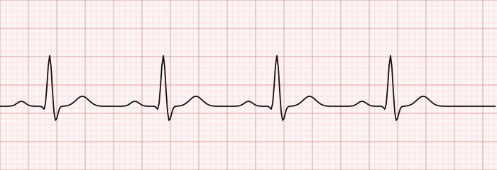
Two close relatives keep the same pattern but change only the rate. Sinus bradycardia is NSR under 60 bpm — often normal in athletes or during sleep, but sometimes a sign of medication effect, high vagal tone, or sinus node disease. Sinus tachycardia is NSR over 100 bpm — usually a response to something (fever, pain, hypovolemia, exercise, anxiety) rather than a primary electrical problem. In both, the defining feature is preserved: one upright P wave before every narrow QRS.
Narrow and Irregular: Atrial Fibrillation and Flutter
When the QRS is narrow but the rhythm is not neatly regular, your attention turns to the atria. Two rhythms dominate this space, and telling them apart is a classic exam distinction.
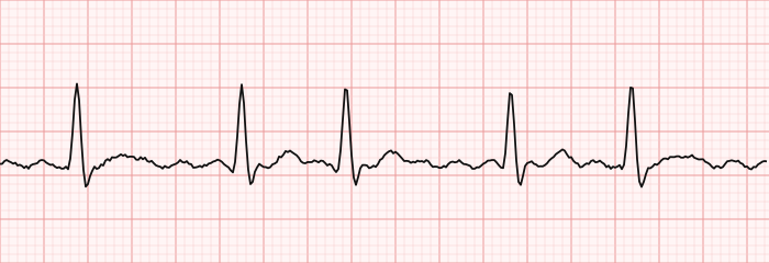
Atrial fibrillation (AFib) is the most common sustained arrhythmia and the archetype of the irregularly irregular rhythm. The organized P waves disappear, replaced by a chaotic, wandering baseline, and the R-R intervals never settle into a pattern. If you march out the QRS complexes and can find no repeating rhythm, AFib is the leading answer. Because the atria quiver rather than contract, blood can pool and clot — which is why AFib is a leading cause of cardioembolic stroke. We cover its mechanism and management in depth on the dedicated atrial fibrillation page.
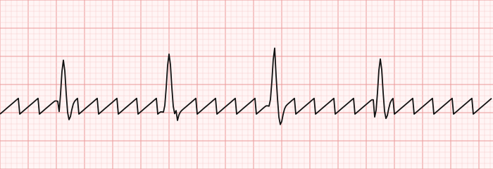
Atrial flutter is the organized cousin. A single reentrant loop, usually circling the right atrium through the cavotricuspid isthmus, fires at a remarkably steady 250–350 bpm (classically ~300), producing the unmistakable sawtooth flutter waves best seen in the inferior leads (II, III, aVF). The AV node lets through only a fraction of these beats, so a 2:1 conduction ratio yields a regular ventricular rate near 150 — a notorious trap that can masquerade as sinus tachycardia. We compare the two rhythms side by side in atrial flutter vs atrial fibrillation.
Narrow and Fast and Regular: SVT
When the QRS is narrow, the rate is fast (often 150–250 bpm), and the rhythm is regular but you cannot identify normal P waves, you are dealing with a supraventricular tachycardia (SVT). The term is a catch-all for fast rhythms originating at or above the AV node, and it includes atrial flutter and AFib — but in everyday use, 'SVT' most often means the reentrant tachycardias: AV nodal reentrant tachycardia (AVNRT) and AV reentrant tachycardia (AVRT).
These rhythms typically start and stop abruptly (paroxysmal), which is why patients describe a sudden 'switch' of the heart racing. The P waves are often buried in the QRS or T wave and hard to see. Because the impulse still travels down the normal His-Purkinje system, the QRS stays narrow — the key feature separating most SVT from ventricular tachycardia.
First-line management of a stable, regular narrow-complex SVT is to interrupt the reentrant loop: vagal maneuvers (Valsalva, carotid sinus massage) first, then adenosine, which briefly blocks the AV node. If the patient is unstable, synchronized cardioversion is used instead. For a fuller treatment, see supraventricular tachycardia; recurrent cases are frequently cured with cardiac ablation of the reentrant pathway.
Wide-Complex Rhythms: VT and VFib
A wide QRS (0.12 s or more) means trouble until proven otherwise, because it signals that the beat is arising in — or being conducted abnormally through — the ventricles. Two ventricular rhythms sit at the dangerous end of the spectrum.
Ventricular tachycardia (VT) is a run of three or more consecutive ventricular beats at a fast rate (usually >100, often 150–200 bpm) with wide, bizarre QRS complexes. Monomorphic VT shows uniform complexes; polymorphic VT varies beat to beat. VT may be tolerated for a short time or may collapse blood pressure entirely, and it can degenerate into ventricular fibrillation. A crucial teaching point: a regular wide-complex tachycardia should be assumed to be VT, especially in a patient with known heart disease, rather than dismissed as SVT with aberrancy. More detail lives on the ventricular tachycardia page.
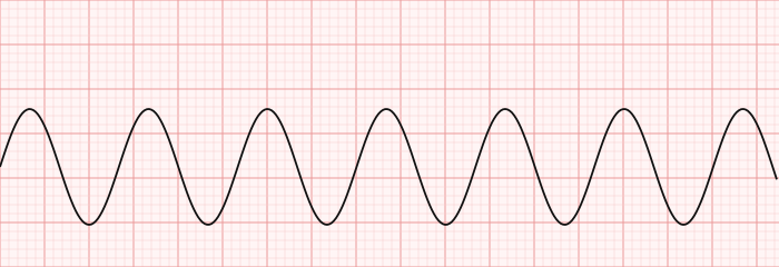
A special polymorphic form, torsades de pointes, twists around the baseline and is linked to a prolonged QT interval — a reason QT monitoring matters, as covered under the QT interval and its bedside calculation via our QTc calculator.
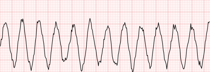
Ventricular fibrillation (VFib) is chaos: no organized QRS complexes at all, just a coarse or fine quivering baseline. The ventricles are not pumping, there is no effective cardiac output, and the patient is in cardiac arrest. VFib is not a rhythm you manage electively — it demands immediate CPR and defibrillation.
AV Heart Block: When P and QRS Lose Sync
Heart block is a failure of the AV node or the conduction system below it to pass atrial impulses to the ventricles reliably. The trick to reading it is to focus on the relationship between P waves and QRS complexes — specifically the PR interval and whether every P is followed by a QRS.
| Type | PR interval | Key feature |
|---|---|---|
| First-degree | Prolonged (>0.20 s), constant | Every P conducts; PR just long. Benign, usually. |
| Second-degree, Mobitz I (Wenckebach) | Progressively lengthens | PR stretches until a QRS is dropped, then resets. Often benign. |
| Second-degree, Mobitz II | Constant, then sudden drop | PR fixed; occasional P waves fail to conduct. Higher risk of progression. |
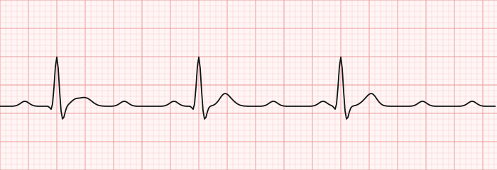
| Third-degree (complete) | No relationship | P waves and QRS march independently (AV dissociation). Escape rhythm drives ventricles. |
In complete (third-degree) heart block, the atria and ventricles beat on entirely separate schedules — the P-P intervals are regular, the R-R intervals are regular, but they have nothing to do with each other. A slow escape rhythm from the AV junction or ventricle keeps the patient alive, but the rate is often dangerously low.
Mobitz II and complete block are the ones that frequently need a pacemaker, because they can progress abruptly to profound bradycardia or asystole. A fuller walkthrough of each grade lives on the heart block page.
Putting It Together: The Narrow vs Wide Decision Tree
Here is the whole framework condensed into one lookup. Start with regularity and QRS width — the two questions from step one — and the plausible rhythms fall out quickly.
| QRS width | Regular | Irregular |
|---|---|---|
| Narrow (<0.12 s) | Sinus rhythm / sinus tach; SVT (AVNRT, AVRT); atrial flutter with fixed conduction | Atrial fibrillation; atrial flutter with variable block; multifocal atrial tachycardia |
| Wide (≥0.12 s) | Ventricular tachycardia; SVT with aberrancy or bundle branch block; paced rhythm | Atrial fibrillation with aberrancy; polymorphic VT / torsades; VFib (no organized QRS) |
Two guardrails keep this tree safe. First, a wide, regular tachycardia is VT until proven otherwise — do not talk yourself into a benign explanation. Second, an irregularly irregular narrow rhythm is AFib until proven otherwise. Those two defaults resolve a large share of real-world strips and exam questions.
Once you can classify the rhythm, you can start reasoning about its hemodynamic impact — how the loss of atrial kick or an extreme rate changes filling and output. That link between electrical and mechanical events is the bridge to our hemodynamics guide, where rhythm meets pressure and flow. For deeper drills, the ECG guide ties the waveform fundamentals to rhythm recognition.
Artifact, Look-Alikes, and Common Traps
A surprising number of 'arrhythmias' are not arrhythmias at all — they are artifact or predictable look-alikes. Training your eye to catch them prevents both exam errors and unnecessary alarm at the bedside.
- Artifact mimicking VT or VFib. A loose electrode, patient tremor (Parkinson's, shivering), or tooth-brushing motion can throw a chaotic tracing that looks like VFib. The giveaway: you can often march out normal QRS complexes through the noise, and the patient is awake and talking.
- Flutter hiding as sinus tach. As noted, 2:1 flutter sits at ~150 bpm and hides its sawtooth inside the QRS-T. Any regular narrow tachycardia at that rate deserves a second look at the inferior leads.
- VT versus SVT with aberrancy. Both are wide and fast. Favor VT with older age, known structural heart disease or prior myocardial infarction, AV dissociation, and very wide/bizarre complexes. When in doubt, treat as VT.
- Paced rhythm. Sharp pacing spikes before the P or QRS reveal a pacemaker at work; do not mistake a paced wide QRS for VT.
Because the same rapid or irregular rhythm distorts intra-arterial and intracardiac pressure tracings, technologists supporting hemodynamic studies need to recognize when the rhythm — not the transducer — is causing an odd waveform. That crossover is part of the day-to-day scope of the cardiovascular ultrasound technologist and is refined by practicing rhythms against pressures in the lab.
How Rhythms Show Up on the RCIS Exam
On the credentialing exam and in the cath lab, rhythm questions reward the same disciplined process this guide teaches. You will be handed a strip and asked to name it, distinguish two look-alikes, or pick the correct first response. Speed comes from the framework, not from cramming pictures.
High-yield points to carry in:
- Irregularly irregular, no P waves = AFib. The single most testable pattern.
- Sawtooth in II/III/aVF = atrial flutter; a regular ~150 narrow tachycardia is 2:1 flutter until proven otherwise.
- Narrow, regular, fast, abrupt onset, no clear P = SVT; treat with vagal maneuvers then adenosine when stable.
- Wide + regular + fast = VT until proven otherwise; never give an AV-nodal blocker to undiagnosed wide-complex tachycardia.
- No organized QRS = VFib — shock and CPR.
- P and QRS dissociated = complete heart block; Mobitz II and complete block trend toward needing a pacemaker.
Drilling real tracings is the fastest way to make these reflexive. Work through the targeted RCIS ECG practice questions, then the ECG strip identification set, which places these rhythms side by side so the contrasts stick. For the acute-ischemia strips that overlap with rhythm reading, our STEMI ECG interpretation guide covers ST-elevation recognition.
Key Takeaways
- Systematize rhythm reading: rate, regularity, QRS width, P-to-QRS relationship, and PR interval — asked in the same order every time.
- Regular or irregular, narrow or wide are the first two questions and route you into the correct diagnostic quadrant before you hunt for P waves.
- Normal sinus rhythm — one upright P before every narrow QRS at 60–100 bpm — is the reference you compare everything against.
- Irregularly irregular with no P waves is atrial fibrillation; regular sawtooth waves are atrial flutter, which loves to hide at ~150 bpm with 2:1 conduction.
- Narrow, regular, fast, abrupt-onset SVT responds to vagal maneuvers and adenosine when stable.
- Any wide, regular tachycardia is ventricular tachycardia until proven otherwise; ventricular fibrillation shows no organized QRS and is a shockable arrest rhythm.
- Heart block is read through the PR interval and the P-QRS relationship; complete block shows fully dissociated P waves and QRS complexes and often needs a pacemaker.
- This is educational content for RCIS preparation, not medical advice; management follows current guidelines and the treating team.
Identify rhythms on real strips
Practise rhythm recognition with instant feedback.
Practise Strips →Frequently asked questions
What are the five steps to interpret an ECG rhythm?
Ask five questions in order: What is the rate? Is the rhythm regular or irregular? Is the QRS narrow or wide? Is there a P wave for every QRS with a consistent relationship? And is the PR interval normal and constant? Answering these in sequence routes almost any strip to the correct rhythm without relying on pattern memory.
How do you tell a narrow-complex rhythm from a wide-complex rhythm?
Measure the QRS duration. A QRS under 0.12 seconds (less than three small boxes) is narrow, meaning the impulse originated at or above the AV node and used the normal conduction system. A QRS of 0.12 seconds or more is wide, indicating the beat arose in the ventricle or was conducted abnormally — which raises the concern for ventricular tachycardia.
What does 'irregularly irregular' mean on an ECG?
It describes R-R intervals that follow no repeating pattern at all — the spacing between beats is random. It is the hallmark of atrial fibrillation, in which organized P waves are absent and replaced by a chaotic baseline. This differs from a 'regularly irregular' rhythm, which repeats a predictable pattern of grouped beats.
How do you distinguish atrial flutter from atrial fibrillation?
Atrial flutter shows regular sawtooth flutter waves, best seen in leads II, III, and aVF, and usually a regular ventricular rate because the AV node conducts in a fixed ratio. Atrial fibrillation has no discrete P waves, a wavering chaotic baseline, and an irregularly irregular ventricular response with no repeating pattern.
Why is a regular wide-complex tachycardia treated as VT until proven otherwise?
Because ventricular tachycardia is the most dangerous and most common cause of a regular wide-complex tachycardia, especially in patients with known heart disease. Mistaking VT for SVT with aberrancy and giving an AV-nodal blocking drug can worsen the rhythm or cause collapse. Assuming VT and treating accordingly is the safe default.
How is ventricular fibrillation recognized on a rhythm strip?
Ventricular fibrillation shows no organized QRS complexes — only a disorganized, quivering baseline that may be coarse or fine. There is no measurable rate, no P waves, and no effective cardiac output. It is a cardiac-arrest rhythm requiring immediate CPR and defibrillation, not an elective rhythm to analyze at leisure.
What is the difference between the types of heart block?
First-degree block has a long but constant PR interval with every P conducting. Second-degree Mobitz I (Wenckebach) shows a progressively lengthening PR until a beat is dropped. Mobitz II drops beats suddenly with a fixed PR. Third-degree (complete) block has P waves and QRS complexes that beat completely independently of each other.
Why does atrial flutter often produce a heart rate near 150 bpm?
In typical atrial flutter the atria fire around 300 beats per minute, and the AV node commonly conducts every other impulse in a 2:1 ratio, giving a ventricular rate near 150. Because the rhythm is regular at that rate, it can be mistaken for sinus tachycardia until the hidden sawtooth flutter waves are unmasked.
How can I tell artifact from a real dangerous rhythm?
Look for normal QRS complexes marching through the noise and check the patient. Artifact from a loose lead, tremor, or movement can mimic ventricular tachycardia or fibrillation, but you can usually find underlying regular beats within the chaos, and the patient remains awake and stable. A true lethal rhythm is matched by clinical collapse.
Sources & further reading
- Cardiovascular Credentialing International (CCI)
- American College of Cardiology
- American Heart Association
- MedlinePlus (U.S. National Library of Medicine)
External links are provided for reference; always confirm current details with the official source.