Atrial Flutter vs Atrial Fibrillation
Atrial flutter and atrial fibrillation are cousins — both are fast, supraventricular rhythms that raise stroke risk — but they look different on the strip, arise from different mechanisms, and respond differently to ablation. Telling them apart is a core RCIS skill and a favorite exam distinction.
- Atrial Flutter vs Atrial Fibrillation: The Quick Distinction
- Different Mechanisms: Reentry Loop vs Chaotic Wavelets
- Reading the ECG: Sawtooth vs Irregular Baseline
- Side-by-Side Comparison Table
- Why the Difference Matters Clinically
- Treatment: Rate Control, Rhythm Control, and Anticoagulation
- Ablation: Where Flutter and Fibrillation Diverge Most
- How Flutter and Fibrillation Coexist and Convert
- On the RCIS Exam and in the Cath Lab
- Key Takeaways
Atrial Flutter vs Atrial Fibrillation: The Quick Distinction
If you strip away every nuance, the difference between these two rhythms comes down to one word: organization. Atrial flutter is an organized atrial arrhythmia — a single, disciplined electrical wave marching around a fixed circuit. Atrial fibrillation is a disorganized one — dozens of chaotic wavelets colliding across the atria with no coordinated beat at all. That single structural difference explains almost everything else: how each looks on the ECG, how regular the pulse feels, and how neatly each responds to catheter ablation.
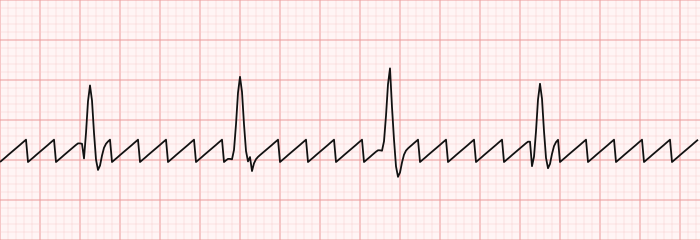
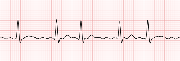
On the surface tracing, atrial flutter produces the classic sawtooth pattern of regular flutter waves, most obvious in the inferior leads (II, III, aVF). Atrial fibrillation, by contrast, replaces organized P waves with a fine, wandering, irregular baseline and — its signature — an irregularly irregular ventricular response. Learning to read both is easier once you are comfortable with the fundamentals in our ECG interpretation primer and the broader survey of ECG rhythm interpretation.
Both belong to the family of supraventricular tachycardias, so a quick review of supraventricular tachycardia helps frame where they sit. And because atrial fibrillation is by far the more common of the two, we cover it in depth on its own dedicated atrial fibrillation page — this article focuses on how the two rhythms differ.
This article is educational and written for RCIS exam preparation. It is not medical advice; clinical decisions belong to the treating team following current guidelines.
Different Mechanisms: Reentry Loop vs Chaotic Wavelets
The clearest way to keep these rhythms straight is to understand what the atrial tissue is actually doing.
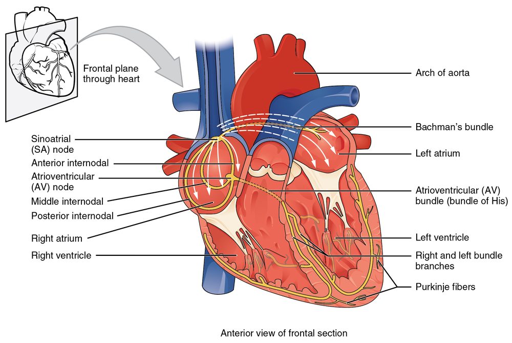
Atrial flutter is a macro-reentrant circuit. In the common (typical) form, a single wave of depolarization travels in a large, fixed loop around the right atrium, usually passing through a narrow strip of tissue called the cavotricuspid isthmus (CTI) — the corridor between the tricuspid valve and the inferior vena cava. Because the circuit is anatomically stable and the wave cycles at a fixed rate, the atria fire at a remarkably consistent 250 to 350 beats per minute. Typical counterclockwise flutter gives the negative sawtooth in the inferior leads; a clockwise circuit reverses that pattern.
Atrial fibrillation is multiple, shifting wavelets. Instead of one tidy loop, AF is driven by rapid, disorganized firing — often triggered by ectopic foci in the pulmonary veins — that fragments into many small reentrant wavelets wandering across both atria. There is no single dominant circuit, so the atria never contract as a unit. Effective atrial rate can exceed 400 to 600 impulses per minute, but these are quivering, not beating.
To picture where these circuits live and why the pulmonary veins matter, it helps to review cardiac structure in our cardiac anatomy guide and the electrical pathway in the cardiac conduction system overview. The AV node acts as a gatekeeper for both rhythms — it cannot conduct every atrial impulse, so it filters that rapid atrial activity down to a ventricular rate the heart can survive.
Reading the ECG: Sawtooth vs Irregular Baseline
This is where the two rhythms declare themselves, and where the RCIS exam most often tests you. Start by asking two questions: Are there organized atrial waves? and Is the ventricular rhythm regular?
Atrial flutter. The hallmark is the sawtooth flutter waves (F waves) — uniform, regular, saw-blade deflections best seen in leads II, III, and aVF. There is no flat, isoelectric baseline between them; the tracing looks continuously toothed. Because the atrial rate is so consistent (about 300 bpm in typical flutter) and the AV node usually conducts in a fixed ratio, the ventricular rate is often regular. A 2:1 block gives a ventricular rate near 150 bpm — a classic exam trap, because at that speed the alternate flutter waves can hide inside the QRS and T waves and masquerade as a plain sinus tachycardia.
Atrial fibrillation. Here the organized P waves vanish entirely, replaced by a fine, chaotic, wandering baseline of fibrillatory (f) waves. The defining fingerprint is the ventricular response: irregularly irregular, with no repeating pattern to the R-R intervals. If you march out the QRS complexes and they never settle into a rhythm, you are almost certainly looking at AF.
Drilling real strips is the fastest way to lock this in — our ECG strip identification set and the broader RCIS ECG practice questions put flutter and fibrillation side by side with other rhythms so the contrast sticks.
Side-by-Side Comparison Table
When you need the difference at a glance, this table captures the features most likely to appear on the exam and at the bedside.
| Feature | Atrial Flutter | Atrial Fibrillation |
|---|---|---|
| Mechanism | Single macro-reentrant loop (often CTI-dependent) | Multiple chaotic wavelets / pulmonary-vein triggers |
| Atrial rate | ~250–350 bpm (classically ~300) | ~400–600 quivering impulses/min |
| Atrial waves | Sawtooth F waves, no flat baseline | No P waves; fine irregular baseline |
| Ventricular rhythm | Often regular (fixed conduction ratio) | Irregularly irregular |
| Best diagnostic leads | II, III, aVF (inferior) | Any; look at R-R irregularity |
| Classic pitfall | 2:1 flutter at ~150 mimics sinus tachycardia | Fine AF mistaken for artifact |
| Ablation success | Very high for typical CTI flutter (~90%+) | Good but lower; often needs pulmonary-vein isolation |
| Stroke risk | Elevated (managed like AF) | Elevated; leading cause of cardioembolic stroke |
Notice that the two rhythms converge on the things that matter most clinically — both drive stroke risk and both demand rate or rhythm control — even as they diverge sharply in appearance and in how cleanly ablation can cure them.
Why the Difference Matters Clinically
These are not just two ECG patterns to memorize. The distinction shapes symptoms, hemodynamics, and treatment strategy.
Loss of the atrial kick. In both rhythms the atria stop contracting effectively, so the heart loses the atrial 'kick' that normally tops off ventricular filling in late diastole — roughly 15 to 30 percent of ventricular filling in a stiff or failing heart. That lost priming volume reduces stroke volume and, at fast rates, drags down cardiac output. Patients with diastolic dysfunction or reduced ejection fraction feel this loss the hardest, which is why new AF or flutter can tip a compensated heart-failure patient into decompensation.
Rate and tachycardia-mediated harm. A poorly controlled ventricular rate over weeks to months can weaken the heart muscle itself — tachycardia-induced cardiomyopathy — a reason rate control is not merely symptomatic. The interplay of rate, filling, and output is worth reviewing in our hemodynamics guide, since these rhythms are a live illustration of the principles.
Stroke risk is the headline. When the atria quiver instead of contract, blood stagnates — especially in the left atrial appendage — and can clot. If a clot embolizes, it can cause a stroke. Crucially, atrial flutter is not the safer rhythm here: its stroke risk is high enough that it is managed with the same anticoagulation thresholds as atrial fibrillation. That equivalence is one of the most important practical takeaways of the whole comparison.
Treatment: Rate Control, Rhythm Control, and Anticoagulation
Management of both rhythms rests on three pillars, and current guidance treats them largely in parallel — though flutter's fixed circuit makes it the more curable of the two.
1. Anticoagulation to prevent stroke. This is decided by patient risk, not by how fast the heart is going. Clinicians use a validated risk score (the CHA2DS2-VASc framework) to weigh factors like heart failure, hypertension, age, diabetes, prior stroke, vascular disease, and sex. When risk crosses the threshold, oral anticoagulation — increasingly a direct oral anticoagulant rather than warfarin — is recommended for flutter and fibrillation alike. This is guideline-driven and individualized; it is educational context here, not a prescription.
2. Rate control. Slowing AV conduction lets the ventricles fill and eases symptoms. Beta-blockers and non-dihydropyridine calcium channel blockers (diltiazem, verapamil) are the usual first-line agents; digoxin is a secondary option, particularly with heart failure. Notably, flutter is often harder to rate-control than AF because its regular, fixed conduction resists pharmacologic slowing.
3. Rhythm control. This means restoring and maintaining sinus rhythm — through electrical or pharmacologic cardioversion, antiarrhythmic drugs, or catheter ablation. Recent evidence has pushed rhythm control, and especially early ablation, toward the front of the line for many symptomatic patients rather than reserving it as a last resort, though the best strategy remains individualized and the evidence continues to evolve.
| Pillar | Goal | Typical tools |
|---|---|---|
| Anticoagulation | Prevent cardioembolic stroke | DOACs or warfarin, guided by risk score |
| Rate control | Slow ventricular response, ease symptoms | Beta-blockers, diltiazem/verapamil, digoxin |
| Rhythm control | Restore/maintain sinus rhythm | Cardioversion, antiarrhythmics, ablation |
Any patient who is hemodynamically unstable — hypotensive, ischemic, or in acute heart failure from the arrhythmia — bypasses this ladder and goes to urgent synchronized cardioversion. If cardioversion is planned and the rhythm has lasted longer than 48 hours (or is of unknown duration), the atrium is first cleared of clot, either with several weeks of anticoagulation or a transesophageal echocardiogram to rule out a left atrial appendage thrombus.
Ablation: Where Flutter and Fibrillation Diverge Most
Nowhere is the difference between these rhythms more consequential than in the electrophysiology lab, because their mechanisms make one far easier to cure than the other. Catheter cardiac ablation destroys the tissue responsible for the arrhythmia — but 'the tissue responsible' is a single narrow corridor in flutter and a diffuse, moving target in fibrillation.
Typical atrial flutter is one of the great success stories of ablation. Because the common form depends on the cavotricuspid isthmus, an electrophysiologist can place a line of ablation lesions across that isthmus and interrupt the entire circuit. This CTI ablation is quick, low-risk, and durable, with single-procedure success rates commonly quoted above 90 percent. For symptomatic typical flutter, ablation is often chosen as a first-line rhythm-control strategy rather than a fallback.
Atrial fibrillation ablation is more involved. Since AF is frequently triggered by the pulmonary veins, the cornerstone procedure is pulmonary vein isolation (PVI) — encircling and electrically disconnecting the veins from the left atrium, using radiofrequency energy, cryoablation, or newer pulsed-field ablation. It works well, but the atrium is a larger and more variable substrate, so success rates are lower than for flutter and repeat procedures are more common, particularly in persistent AF or an enlarged left atrium. Even so, guidelines increasingly support offering ablation early to appropriate symptomatic patients.
An important clinical wrinkle ties the two together: many patients have both rhythms, and some develop atrial flutter after AF ablation or while on antiarrhythmic drugs. Understanding both circuits — and how a diagnostic study in a cardiac cath lab or EP lab maps them — is part of the complete picture.
| Ablation feature | Typical Atrial Flutter | Atrial Fibrillation |
|---|---|---|
| Target | Cavotricuspid isthmus (one line) | Pulmonary vein isolation (encircling) |
| Complexity | Lower, shorter procedure | Higher, longer procedure |
| Single-procedure success | Very high (~90%+) | Good; often needs repeat, esp. persistent AF |
| First-line consideration | Frequently first-line for typical flutter | Increasingly early, still individualized |
How Flutter and Fibrillation Coexist and Convert
It is tempting to treat these as two separate diseases, but in practice they live on a continuum and frequently swap back and forth. The same atrial disease — stretch, fibrosis, inflammation, prior surgery — that fosters one predisposes to the other. A patient can be in flutter one day and fibrillation the next, and a monitor may capture both on the same recording.
Several patterns are worth knowing for the exam and the lab:
- AF degenerating from flutter, and vice versa. Organized flutter can break down into fibrillation as wavelets fragment; conversely, treated or partially organized AF can settle into a flutter circuit.
- Drug-induced 'hybrid' flutter. Antiarrhythmic drugs used for AF can slow atrial conduction just enough to organize the rhythm into an atrial flutter — sometimes then treated by adding CTI ablation.
- Post-ablation flutter. After AF ablation, scar lines can create new reentrant circuits (atypical, or 'scar-related,' flutters) that behave like flutter but sit outside the classic isthmus.
Both rhythms also share the same broad risk factors — hypertension, valvular disease, obstructive sleep apnea, obesity, hyperthyroidism, and structural heart disease from coronary artery disease or prior myocardial infarction. Managing those upstream drivers is now considered part of rhythm management, not just an afterthought.
For the cardiovascular technologist, the takeaway is to read the strip rhythm by rhythm rather than assuming a single diagnosis holds for a whole study — and to recognize that stroke prophylaxis follows the patient's risk profile regardless of which of the two rhythms is showing at any given moment.
On the RCIS Exam and in the Cath Lab
For the registered cardiovascular invasive specialist, this comparison shows up in two places: on the credentialing exam and on the live monitor during procedures. On the exam, expect to be handed a rhythm strip and asked to name it, to distinguish flutter from fibrillation from other supraventricular tachycardias, and to identify the correct first-line management concept.
High-yield points to carry into the test:
- Sawtooth = flutter; irregularly irregular with no P waves = fibrillation. That single pair resolves most questions.
- A regular narrow-complex tachycardia at ~150 bpm should trigger suspicion for 2:1 atrial flutter.
- Both carry stroke risk and use the same anticoagulation logic — flutter is not the 'safe' one.
- Typical flutter ablates at the cavotricuspid isthmus with very high success; AF ablation centers on pulmonary vein isolation.
In the procedure room, both rhythms can appear during hemodynamic studies and interventions. A tech who supports pressure measurements should recognize how an irregular or rapid rhythm distorts waveform interpretation — a topic that connects to the fundamentals in our hemodynamics guide and to the day-to-day scope of the cardiovascular ultrasound technologist role. To sharpen recognition under exam-style pressure, run through the targeted RCIS ECG practice set alongside the strip drills.
Key Takeaways
- The core difference: atrial flutter is one organized reentrant loop; atrial fibrillation is many chaotic wavelets with no coordinated atrial contraction.
- On the ECG, flutter shows regular sawtooth waves (best in II, III, aVF), while fibrillation shows no P waves and an irregularly irregular ventricular response.
- Beware the ~150 bpm regular narrow tachycardia — it is often 2:1 atrial flutter masquerading as sinus tachycardia.
- Both rhythms lose the atrial kick, reduce cardiac output at fast rates, and carry a serious stroke risk managed with the same anticoagulation logic.
- Treatment rests on three pillars: anticoagulation by risk score, rate control, and rhythm control.
- Ablation diverges sharply: typical flutter is cured by a single cavotricuspid-isthmus line (~90%+ success), whereas fibrillation needs pulmonary vein isolation and more often a repeat procedure.
- The two rhythms coexist and convert into one another, sharing risk factors and often the same patient.
- This is educational content for RCIS preparation, not medical advice.
Frequently asked questions
What is the main difference between atrial flutter and atrial fibrillation?
Atrial flutter is an organized rhythm driven by a single reentrant loop, producing regular sawtooth flutter waves and often a regular ventricular rate. Atrial fibrillation is disorganized, driven by many chaotic wavelets, producing no true P waves and an irregularly irregular pulse. In short: flutter is ordered, fibrillation is chaotic.
How do you tell atrial flutter from atrial fibrillation on an ECG?
Look at the atrial activity and the R-R intervals. Atrial flutter shows uniform sawtooth flutter waves, best seen in leads II, III, and aVF, and usually a regular ventricular rhythm. Atrial fibrillation has no discrete P waves, a wavering baseline, and an irregularly irregular ventricular response with no repeating pattern.
Is atrial flutter more dangerous than atrial fibrillation?
Neither is clearly 'safer.' Atrial flutter looks more organized, but its stroke risk is high enough that it is anticoagulated using the same risk-based thresholds as atrial fibrillation. Both can also cause rapid heart rates and reduce cardiac output, so both are treated seriously.
Why does atrial flutter often cause a heart rate around 150 bpm?
In typical atrial flutter the atria fire at roughly 300 beats per minute, and the AV node commonly conducts every other impulse — a 2:1 ratio — giving a ventricular rate near 150 bpm. Because the rhythm is regular at that rate, it can be mistaken for sinus tachycardia until the hidden sawtooth waves are unmasked.
Can atrial flutter turn into atrial fibrillation?
Yes. The two rhythms share the same underlying atrial disease and frequently convert into one another. Organized flutter can fragment into fibrillation, and fibrillation can organize into a flutter circuit, sometimes after antiarrhythmic drugs or ablation. Many patients experience both over time.
Is ablation more successful for atrial flutter or atrial fibrillation?
Ablation for typical atrial flutter is more successful because it targets a single, fixed corridor — the cavotricuspid isthmus — with single-procedure success commonly above 90 percent. Atrial fibrillation ablation centers on isolating the pulmonary veins, works well but has lower success rates, and more often requires a repeat procedure.
Do both atrial flutter and atrial fibrillation require blood thinners?
Anticoagulation is decided by a patient's stroke-risk profile rather than by which of the two rhythms is present. Both flutter and fibrillation use the same risk-scoring framework, so a patient who meets the threshold is generally recommended anticoagulation regardless of whether they are in flutter or fibrillation. This is individualized, guideline-driven care.
What is the sawtooth pattern on an ECG?
The sawtooth pattern is the classic appearance of atrial flutter: regular, uniform flutter waves that resemble the teeth of a saw blade, with no flat baseline between them. It is most visible in the inferior leads (II, III, and aVF) and reflects the single organized reentrant circuit driving the rhythm.
Why do atrial fibrillation and flutter reduce cardiac output?
Both rhythms stop the atria from contracting effectively, so the heart loses the atrial kick that normally completes ventricular filling. Combined with a fast, sometimes irregular ventricular rate that shortens filling time, this lowers stroke volume and cardiac output — an effect felt most by patients with stiff or weakened hearts.
Sources & further reading
- Cardiovascular Credentialing International (CCI)
- American College of Cardiology
- American Heart Association
- MedlinePlus (U.S. National Library of Medicine)
External links are provided for reference; always confirm current details with the official source.