Cardiac Ablation: How It Works & What to Expect
Cardiac ablation is a catheter-based procedure that deliberately destroys the tiny patch of heart tissue driving an abnormal rhythm — and for arrhythmias like AFib, SVT, and VT it has moved from last resort to front-line therapy.
- What Is Cardiac Ablation?
- How Catheter Ablation Works
- Ablation Energy Sources: RF, Cryo, and PFA
- Pulmonary Vein Isolation and Ablation for AFib
- Ablation for SVT and Atrial Flutter
- Ablation for Ventricular Tachycardia
- Risks, Recovery, and Success Rates
- Ablation vs Medication and Devices
- Cardiac Ablation on the RCIS Exam
- Key Takeaways
What Is Cardiac Ablation?
Cardiac ablation is a minimally invasive treatment for heart rhythm disorders. Using thin, steerable catheters threaded through a vein or artery into the heart, an electrophysiologist locates the exact tissue responsible for an arrhythmia and then delivers energy to that spot to create a small, controlled scar. Because scarred tissue does not conduct electricity, the abnormal signal is either destroyed at its source or fenced off so it can no longer trigger or sustain the rhythm.
The word you will hear most often is catheter ablation — a reminder that the whole thing is done from inside the vessels and chambers, without opening the chest. A related but separate approach, surgical ablation (such as the Cox-Maze procedure), achieves the same goal with an open or minimally invasive operation, but the catheter-based version is by far the more common and the one most relevant to the RCIS exam and the electrophysiology (EP) lab.
Ablation does not fix the heart's pump or its arteries; it targets the electrical problem. To understand what it is correcting, it helps to be fluent with the normal wiring described in our overview of the cardiac conduction system and the chamber anatomy in our cardiac anatomy guide. When the SA node, AV node, and His-Purkinje network are working normally, one orderly impulse sweeps the heart per beat. Ablation is what you reach for when a stray focus or an extra circuit hijacks that orderly sequence.
This article is educational and written for RCIS exam preparation. It is not medical advice; treatment decisions belong to the patient and their care team following current guidelines.
How Catheter Ablation Works
A catheter ablation is performed in the EP lab, a cousin of the standard cardiac cath lab equipped with specialized mapping and recording systems. The operator gains access — most commonly through the femoral vein in the groin — and advances catheters under fluoroscopic and, increasingly, three-dimensional electroanatomic guidance into the correct chamber. For left-sided targets like the pulmonary veins, a transseptal puncture is used to cross from the right atrium into the left atrium.
The core steps are consistent across arrhythmia types:
- Access and catheter placement — venous (and sometimes arterial) sheaths are placed, and diagnostic and ablation catheters are positioned in the heart.
- Induction and mapping — the team provokes the arrhythmia if needed and uses recording catheters and 3D mapping to pinpoint the origin or circuit. This electrical detective work is what the whole procedure hinges on.
- Ablation — energy is delivered to the target tissue to create the lesion.
- Confirmation — the operator retests to confirm the arrhythmia can no longer be induced and, where relevant, that conduction is blocked across the ablation line.
Familiarity with the EP lab overlaps heavily with the invasive skills tested on the credential. Techs comfortable with vascular access, catheter handling, and rhythm monitoring — the same competencies covered in our ECG guide — adapt quickly to the EP environment. Understanding how the cardiovascular ultrasound technologist role supports imaging, especially intracardiac echocardiography during transseptal puncture, rounds out the team picture.
Ablation Energy Sources: RF, Cryo, and PFA
The energy used to make the lesion has evolved substantially, and the RCIS exam increasingly expects familiarity with more than one modality.
Radiofrequency (RF) ablation is the classic workhorse. Alternating current heats the catheter tip, and resistive heating of the adjacent tissue raises it to a temperature that produces coagulation necrosis — a controlled thermal burn. RF is versatile and point-by-point, letting the operator draw precise lines and target discrete foci.
Cryoablation does the opposite: it freezes tissue to destroy it. A balloon or focal catheter cools to well below freezing, forming an ice lesion. The cryoballoon is especially popular for pulmonary vein isolation because a single balloon inflation can encircle a vein in one shot. Cryo also allows 'cryomapping' — a reversible cooling test before a permanent lesion, useful near delicate structures like the AV node.
Pulsed field ablation (PFA) is the newer, rapidly expanding option. Instead of heat or cold, PFA delivers short, high-voltage electrical pulses that create pores in cell membranes — a process called irreversible electroporation. Its major appeal is tissue selectivity: cardiac muscle is more susceptible than surrounding structures, which appears to reduce the risk of collateral damage to the esophagus and phrenic nerve. Evidence is still maturing, but PFA has moved quickly into mainstream AFib practice.
| Energy | Mechanism | Typical use | Notable feature |
|---|---|---|---|
| Radiofrequency | Heat (coagulation necrosis) | SVT, VT, AFib, atrial flutter | Precise, point-by-point lesions |
| Cryoablation | Freezing (ice lesion) | PVI for AFib, AVNRT | Single-shot balloon; reversible cryomapping |
| Pulsed field (PFA) | Electroporation (non-thermal) | PVI for AFib | Tissue-selective; less collateral injury |
Pulmonary Vein Isolation and Ablation for AFib
Atrial fibrillation is the single most common reason patients undergo ablation, and pulmonary vein isolation (PVI) is the cornerstone of the procedure. The insight behind it, established decades ago, is that most triggering ectopic beats in paroxysmal AFib originate from muscle sleeves inside the pulmonary veins where they enter the left atrium. Electrically isolating those veins — drawing continuous ablation lines around them so their chaotic impulses can no longer reach the atrium — removes the trigger.
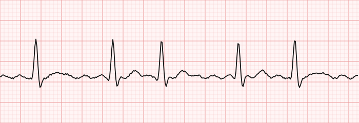
Current guidance has steadily elevated the role of catheter ablation for AFib. For symptomatic paroxysmal AFib, ablation is now a reasonable first-line rhythm-control strategy in appropriately selected patients, rather than something reserved for after drugs fail. It is generally more effective than antiarrhythmic medication at maintaining sinus rhythm and improving symptoms. In patients with AFib and heart failure with reduced ejection fraction, ablation has also been associated with meaningful outcome benefits — an area where the evidence has grown notably strong.
For persistent AFib, PVI remains the foundation, but achieving durable rhythm control is harder, and operators may add lesions beyond the veins. The overall picture is still evolving, and success is best understood as improved rhythm control and quality of life rather than a guaranteed permanent cure. If you want the underlying rhythm fundamentals, our dedicated page on atrial fibrillation and the broader ECG rhythm interpretation guide cover how AFib looks and behaves.
Ablation for SVT and Atrial Flutter
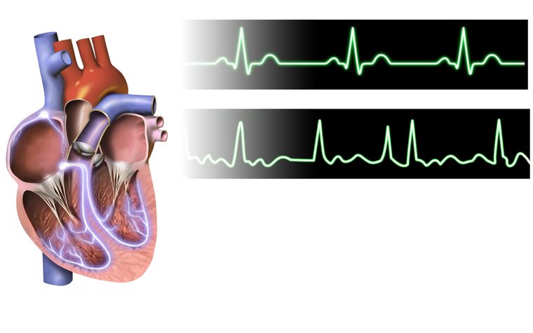
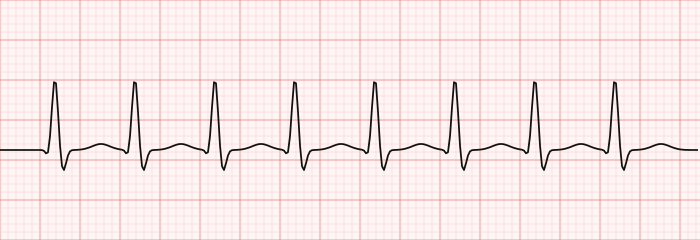
If AFib is the highest-volume target, the supraventricular tachycardias are the most curable. For many forms of supraventricular tachycardia (SVT), catheter ablation is highly effective — success rates frequently exceed 95% — and is often preferred over lifelong medication, especially in young, otherwise healthy patients.
The two classic reentrant SVTs are neatly suited to ablation because they depend on a discrete, targetable pathway:
- AVNRT (AV nodal reentrant tachycardia) uses a small reentry circuit within the AV node region. Ablation modifies the 'slow pathway' to break the loop while sparing normal conduction.
- AVRT (AV reentrant tachycardia), including Wolff-Parkinson-White syndrome, uses an accessory pathway — an extra electrical connection between atrium and ventricle. Ablating that pathway can be definitively curative.
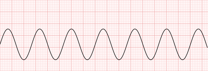
Typical atrial flutter is another textbook ablation success. The rhythm depends on a large reentry circuit circling the right atrium through a narrow corridor called the cavotricuspid isthmus. A single line of ablation across that isthmus blocks the circuit, and the cure rate is excellent. Recognizing flutter's characteristic sawtooth pattern is a common exam task; our ECG interpretation primer and the strip-identification drills below reinforce it.
Ablation for Ventricular Tachycardia
Ablation for ventricular tachycardia (VT) is the most technically demanding branch of the field. VT most often arises from reentry around scar left behind by a prior myocardial infarction or from cardiomyopathy, so the target tissue sits within a diseased, sometimes hard-to-reach ventricle. Mapping identifies the slow-conducting channels through the scar, and ablation interrupts them.
The role of VT ablation has grown. For patients with recurrent monomorphic VT — particularly those getting repeated shocks from an implantable cardioverter-defibrillator (ICD) — ablation reduces arrhythmia burden and shock frequency, and current guidance supports it as an important, sometimes early, option rather than a final measure. It is important to be clear about scope, though: in patients with structural heart disease, ablation generally controls VT and improves quality of life, but most still need an ICD, because ablation lowers the frequency of episodes rather than guaranteeing none will recur.
These patients are often the sickest in the EP lab, and hemodynamic support is a real consideration. Understanding how a failing ventricle behaves ties directly into our hemodynamics guide and the concept of ejection fraction, and unstable cases may involve mechanical support such as the intra-aortic balloon pump. Idiopathic VT in a structurally normal heart — for example, outflow-tract VT — is a happier story, with high ablation success and a generally benign course.
Risks, Recovery, and Success Rates
Catheter ablation is generally safe, but it is a real invasive procedure and carries a small, modality- and target-dependent risk of complications. Being able to name them is a common exam expectation and part of informed patient care.
- Vascular access complications — bleeding, hematoma, or pseudoaneurysm at the groin puncture, the most frequent issue overall.
- Cardiac tamponade — bleeding into the pericardial sac from perforation, which can compress the heart. Recognizing its physiology is covered in our page on cardiac tamponade hemodynamics.
- Heart block — inadvertent injury to the AV node during ablation near it, occasionally requiring a pacemaker. Review the spectrum in our overview of heart block.
- Stroke or thromboembolism — a risk of left-sided procedures, mitigated by anticoagulation.
- Pulmonary vein stenosis, phrenic nerve injury, and (rarely) atrioesophageal fistula — specific to left atrial/PVI work; the atrioesophageal complication is rare but serious.
Recovery from a straightforward catheter ablation is usually quick. Many patients go home the same day or after one night, with a few days of restricted activity to let the access site heal. It is common to feel occasional skipped beats or brief runs of arrhythmia during the first few weeks — the so-called blanking period — as the ablated tissue heals and inflammation settles. Doctors typically wait about three months before judging whether the procedure has succeeded.
Success rates vary widely by rhythm. Reentrant SVT and typical flutter ablations are among the most reliable in all of cardiology. AFib success is good but lower and sometimes requires a repeat procedure, particularly for persistent AFib. VT ablation meaningfully reduces episodes but is best framed as control in a high-risk population.
Ablation vs Medication and Devices
Ablation does not exist in a vacuum — it is one of three broad tools for arrhythmia management, and the exam often probes how they differ.
Antiarrhythmic drugs suppress abnormal rhythms chemically but must be taken indefinitely, carry side effects, and can themselves be proarrhythmic. For many patients ablation offers a chance to reduce or stop these medications. The drugs used acutely in the lab are catalogued in our cath-lab medications reference.
Rhythm devices address a different problem. A pacemaker treats a heart that is too slow; an ICD terminates a lethal ventricular rhythm after it starts. Neither prevents the arrhythmia the way ablation aims to. In fact, the two often work together — VT ablation is frequently done in addition to an ICD to cut down on painful shocks.
| Approach | What it does | Best suited for |
|---|---|---|
| Catheter ablation | Destroys the arrhythmia source/circuit | SVT, flutter, AFib, recurrent VT |
| Antiarrhythmic drugs | Suppress the rhythm pharmacologically | Ongoing control; when ablation is deferred |
| Pacemaker | Paces a slow heart | Bradycardia, heart block |
| ICD | Shocks lethal ventricular rhythms | Sudden-death prevention (often alongside ablation) |
The right choice depends on the arrhythmia, symptoms, patient preference, and comorbidities. A young patient with WPW may be cured outright by ablation, while an older patient with persistent AFib and heart failure might benefit from ablation, a device, and medication in combination. There is rarely one universal answer — which is exactly the nuance well-written exam questions test.
Cardiac Ablation on the RCIS Exam
For the registered cardiovascular invasive specialist, ablation sits at the intersection of electrophysiology, anatomy, and invasive technique. Even if your primary work is in the diagnostic or interventional cath lab, the credential expects you to understand what happens in the EP suite and why.
Expect questions that ask you to match an arrhythmia to its ablation target — pulmonary veins for AFib triggers, the cavotricuspid isthmus for typical flutter, the slow pathway for AVNRT, an accessory pathway for WPW, and scar-related channels for VT. Expect to distinguish the energy modalities (RF heat, cryo cold, PFA electroporation) and to recognize the major complications, especially tamponade and inadvertent heart block. A firm grip on the underlying rhythms — how SVT, flutter, AFib, and VT look on a strip — makes these questions far easier.
To build that fluency, drill our targeted RCIS ECG practice questions and the ECG strip identification set, which include the very rhythms ablation is designed to treat. Pairing rhythm recognition with an understanding of the procedure that fixes it is the fastest way to lock in this domain.
Key Takeaways
- Cardiac ablation destroys the small patch of tissue driving an arrhythmia by creating a controlled scar, done from inside the heart via catheter ablation.
- Energy sources are radiofrequency (heat), cryoablation (cold), and pulsed field ablation (non-thermal electroporation, notable for sparing nearby tissue).
- Pulmonary vein isolation is the cornerstone of AFib ablation, targeting the vein triggers; ablation is now a reasonable first-line rhythm-control option for many patients with symptomatic paroxysmal AFib.
- Reentrant SVTs (AVNRT, AVRT/WPW) and typical atrial flutter have very high ablation cure rates and depend on discrete, targetable circuits.
- VT ablation targets scar-related reentry, reduces ICD shocks, but usually complements rather than replaces an ICD in structural heart disease.
- Main risks include vascular access problems, cardiac tamponade, heart block, and — for left-sided work — stroke and rare pulmonary vein or esophageal injury.
- Recovery is usually quick with a roughly three-month blanking period before judging success.
- This is educational content for RCIS preparation, not medical advice.
Practise the arrhythmias ablation treats
Test AFib, flutter, SVT, and VT recognition.
Practise ECG →Frequently asked questions
What is cardiac ablation and how does it work?
Cardiac ablation is a minimally invasive procedure that treats abnormal heart rhythms by destroying the tissue causing them. Thin catheters are threaded through a vein or artery into the heart, the arrhythmia's source or circuit is mapped, and energy is applied to create a small scar. Because scar does not conduct electricity, the abnormal signal is eliminated or blocked.
What is pulmonary vein isolation?
Pulmonary vein isolation (PVI) is the core step in ablation for atrial fibrillation. Most AFib triggers come from muscle sleeves inside the pulmonary veins where they join the left atrium. PVI draws continuous ablation lines around those veins to electrically isolate them, so their chaotic impulses can no longer reach the atrium and start AFib.
Is cardiac ablation a permanent cure for AFib?
For many patients ablation greatly improves rhythm control and symptoms, and it is more effective than medication at maintaining normal rhythm — but it is not guaranteed to be permanent. Paroxysmal AFib responds better than persistent AFib, and some patients need a repeat procedure. Success is best viewed as durable improvement rather than an absolute cure.
What is the difference between radiofrequency, cryo, and pulsed field ablation?
Radiofrequency ablation uses heat to burn the target tissue point-by-point. Cryoablation freezes tissue to destroy it and is popular for single-shot balloon isolation of the pulmonary veins. Pulsed field ablation is non-thermal, using short high-voltage pulses to porate cell membranes; its main advantage is greater selectivity for cardiac tissue, reducing collateral damage to nearby structures.
Which arrhythmias can be treated with ablation?
Ablation is used for many rhythm disorders, including atrial fibrillation, typical atrial flutter, several forms of supraventricular tachycardia (such as AVNRT and AVRT/Wolff-Parkinson-White), and ventricular tachycardia. Reentrant SVTs and typical flutter have especially high cure rates because they rely on a discrete, targetable circuit.
What are the risks of cardiac ablation?
The most common issues are vascular access complications like bleeding or hematoma at the groin. More serious but less frequent risks include cardiac tamponade from perforation, inadvertent heart block that may need a pacemaker, stroke with left-sided procedures, and — specific to left atrial ablation — pulmonary vein stenosis, phrenic nerve injury, and the rare but serious atrioesophageal fistula.
How long does recovery from catheter ablation take?
Recovery is usually quick. Many patients go home the same day or after one night and resume normal activity within a few days once the access site heals. There is a roughly three-month blanking period during which occasional skipped beats or brief arrhythmias are common as the tissue heals, so doctors typically wait about three months to judge success.
Does ablation replace the need for an ICD in VT?
Usually not in patients with structural heart disease. VT ablation reduces how often episodes occur and cuts down on ICD shocks, which improves quality of life, but it does not reliably prevent every episode. For that reason ablation is generally done alongside an implantable cardioverter-defibrillator rather than as a replacement for it.
Is ablation better than taking antiarrhythmic medication?
For many arrhythmias, especially reentrant SVT and symptomatic paroxysmal AFib, ablation is more effective than antiarrhythmic drugs at maintaining normal rhythm and can reduce or eliminate the need for long-term medication. Drugs remain a valid option, but they must be taken indefinitely, carry side effects, and can occasionally provoke arrhythmias themselves. The best choice depends on the specific rhythm and patient.
Sources & further reading
- Cardiovascular Credentialing International (CCI)
- American College of Cardiology
- American Heart Association
- MedlinePlus (U.S. National Library of Medicine)
External links are provided for reference; always confirm current details with the official source.