Myocardial Infarction (Heart Attack): Types, ECG & Treatment
A myocardial infarction — a heart attack — happens when blood flow to part of the heart muscle is choked off long enough that the tissue begins to die. It is one of the most time-critical emergencies in medicine, where the phrase "time is muscle" is literally true.
What is a myocardial infarction?
A myocardial infarction (MI) is the death of heart muscle caused by a sudden, prolonged loss of its blood supply. The word breaks down cleanly: myo (muscle), cardial (heart), and infarction (tissue death from lack of oxygen). Strip away the jargon and it is exactly what most people mean when they say heart attack.
The heart, despite pumping blood to the entire body, cannot feed itself from the blood inside its chambers. It relies on its own dedicated plumbing — the coronary arteries — that branch across its surface and dive into the muscle. When one of those arteries is abruptly blocked, everything downstream is cut off from oxygen. Within minutes the affected muscle starts to malfunction, and if flow is not restored, cells begin to die in a wave that spreads outward over the following hours.
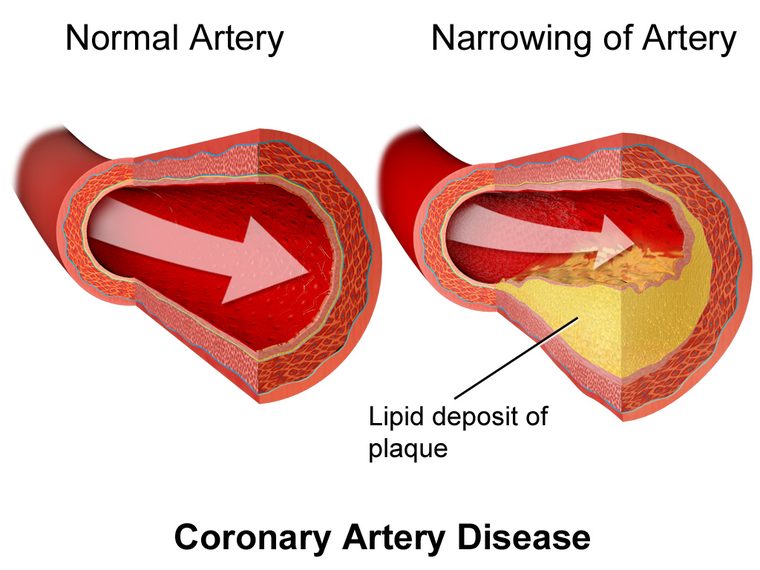
The overwhelming majority of heart attacks are caused by coronary artery disease — years of cholesterol-rich plaque building up inside the artery walls. The trigger for an actual MI is usually not slow narrowing but a sudden event: a plaque ruptures, the body treats it like an injury, and a blood clot forms on top and seals the vessel shut. This is why a person can feel completely well one moment and be in the middle of a heart attack the next.
This article is educational and written for students and clinicians preparing for exams such as the RCIS — it is not medical advice. If you think you or someone near you is having a heart attack, call emergency services immediately.
How a heart attack actually happens
Understanding the mechanism makes everything else — the symptoms, the ECG changes, the treatment — click into place. A heart attack is a chain of events, and each link matters.
It usually begins with a plaque that has been quietly growing inside a coronary artery for years. Not all plaques are equal. The dangerous ones are not necessarily the biggest; they are the ones with a thin, inflamed cap over a soft, fatty core. Under stress — a surge of blood pressure, physical exertion, or sometimes for no obvious reason at all — that cap tears. This is called plaque rupture.
The moment the cap ruptures, the body's clotting system reads it as a wound and rushes to seal it. Platelets pile onto the exposed core and a thrombus (blood clot) forms. If the clot only partly blocks the vessel, flow is throttled but not stopped — this tends to produce an NSTEMI. If the clot completely occludes the artery, the muscle downstream loses its entire supply — this is the classic STEMI. The anatomy of which vessel is involved matters enormously, which is why understanding coronary artery anatomy is central to reading any heart attack.
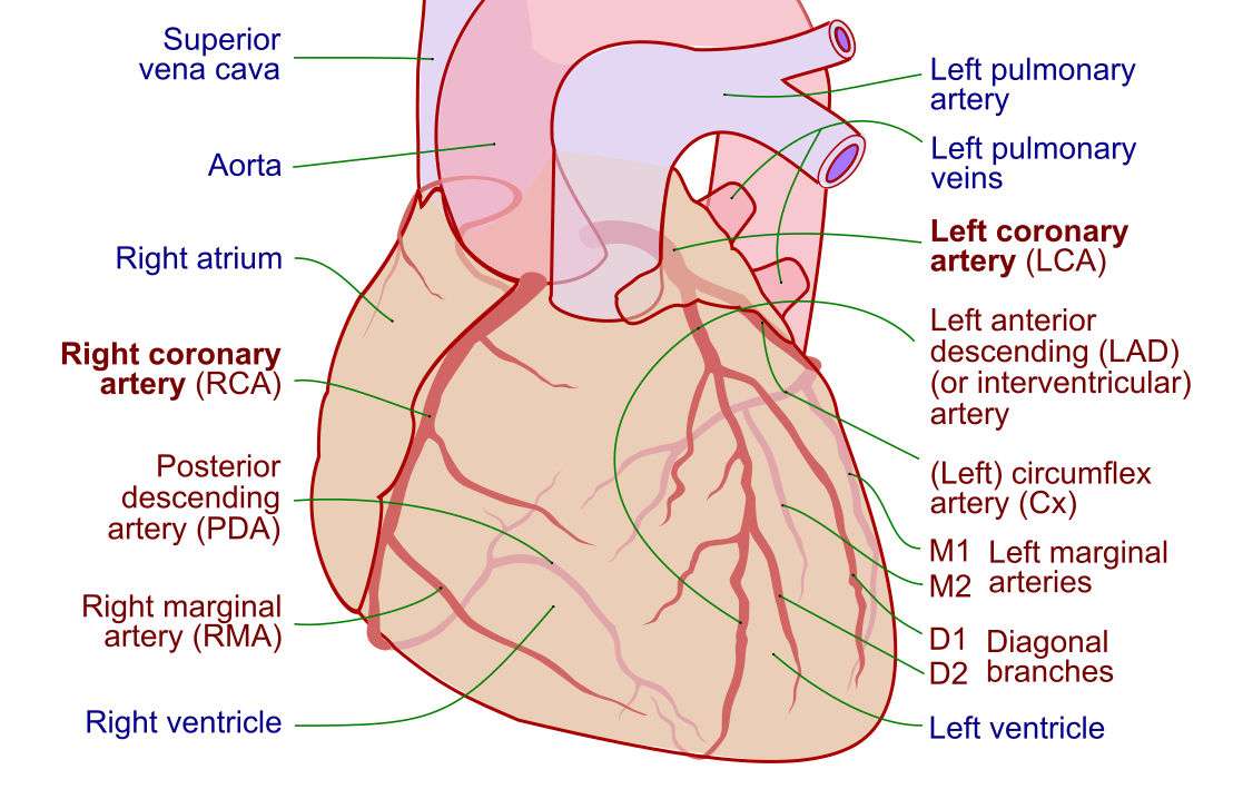
Once flow stops, the clock starts. Deprived of oxygen, heart cells switch to inefficient anaerobic metabolism and begin to fail within minutes. Irreversible death of muscle starts around 20 to 30 minutes into a complete occlusion and marches outward from the innermost layer of the wall toward the outer surface. Restore flow early and much of that muscle can still be saved; wait too long and the damage becomes permanent scar. That biology is the entire reason emergency heart attack care is built around speed.
STEMI vs NSTEMI: the key distinction
If there is one comparison every RCIS candidate must know cold, it is STEMI vs NSTEMI. These two forms of heart attack look similar to the patient but demand different urgency and different treatment pathways. The difference is defined by the electrocardiogram and by how completely the artery is blocked.
STEMI stands for ST-Elevation Myocardial Infarction. The coronary artery is completely blocked, so the full thickness of the heart wall in that territory is starving. On the ECG this produces a hallmark finding: elevation of the ST segment. A STEMI is the most time-critical form — the goal is to reopen the artery as fast as humanly possible.
NSTEMI stands for Non-ST-Elevation Myocardial Infarction. Here the artery is severely narrowed or partially blocked rather than fully occluded, so muscle is being damaged but usually not through the entire wall thickness. The ST segment is not elevated — instead you may see ST depression or T-wave inversion, or the ECG can look nearly normal. The diagnosis leans heavily on blood tests showing that cardiac muscle proteins have leaked into the bloodstream.
| Feature | STEMI | NSTEMI |
|---|---|---|
| Artery blockage | Complete occlusion | Partial / severe narrowing |
| Wall involvement | Full thickness (transmural) | Usually partial thickness |
| ECG hallmark | ST-segment elevation | ST depression, T-wave inversion, or normal |
| Troponin | Elevated | Elevated |
| First-line treatment | Emergency reperfusion (usually PCI) | Risk stratify, then angiography ± PCI |
| Urgency | Immediate — minutes matter | Urgent, but risk-guided timing |
Both STEMI and NSTEMI are "real" heart attacks — both involve dying muscle and both are dangerous. The distinction is not about severity but about strategy. A STEMI means "the pipe is fully shut, open it now." An NSTEMI means "muscle is being injured, stabilize the patient and get to the artery on a risk-guided timeline." A third category, unstable angina, sits alongside NSTEMI in the umbrella of acute coronary syndromes: the same partial-blockage picture but without a rise in cardiac blood markers, meaning ischemia without confirmed muscle death.
Heart attack symptoms
The textbook picture of heart attack symptoms is crushing chest pain — but real life is messier, and knowing the range of presentations saves lives. The classic complaint is a pressure, tightness, squeezing, or heaviness in the center or left side of the chest, often described as "an elephant sitting on my chest" rather than a sharp stab.
That discomfort frequently does not stay put. It radiates — most commonly to the left arm, but also to the jaw, neck, back, or upper abdomen. Alongside the chest discomfort, people often report a cluster of accompanying symptoms:
- Shortness of breath, sometimes as the main or only complaint
- Cold sweat (diaphoresis) — a drenching, clammy sweat
- Nausea or vomiting, easily mistaken for indigestion
- Lightheadedness or a feeling of impending doom
- Unusual fatigue, which can precede the event by days in some people
Critically, not everyone gets the dramatic version. Atypical presentations are common in women, older adults, and people with diabetes, whose nerve signaling may be blunted. They may have no chest pain at all — instead presenting with breathlessness, profound fatigue, jaw or back discomfort, or simply feeling unwell. A "silent" MI can even be discovered later on a routine ECG, having caused symptoms mild enough to be brushed off at the time.
Timing matters as much as the symptoms themselves. Discomfort that comes on with exertion and eases with rest points toward stable angina; discomfort that arrives at rest, is new, or is worsening suggests an acute coronary syndrome and should never be waited out at home.
Reading the ECG in a heart attack
The 12-lead ECG is the single most important early test in a suspected heart attack, and it is fast, cheap, and available anywhere. It is what separates a STEMI from an NSTEMI within minutes of a patient arriving. Building a solid foundation in ECG interpretation is therefore essential for anyone working in cardiovascular care.
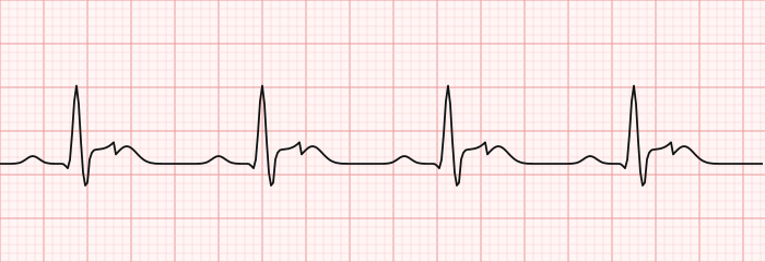
In a STEMI, the giveaway is elevation of the ST segment in leads that face the injured wall. Just as telling, the ECG points to where the infarct is by which leads are affected — and that maps back onto coronary anatomy. A deeper walkthrough lives in our guide to STEMI ECG interpretation, but the core pattern is worth memorizing:
| Location | ECG leads | Usual artery |
|---|---|---|
| Inferior | II, III, aVF | Right coronary artery |
| Anterior / septal | V1–V4 | Left anterior descending |
| Lateral | I, aVL, V5–V6 | Left circumflex |
In an NSTEMI, there is no ST elevation. Instead you may see ST-segment depression, T-wave inversion, or a tracing that looks unremarkable — which is exactly why blood tests are indispensable here. Because the ECG can evolve, a single normal tracing never fully clears a patient with a convincing story; serial ECGs and repeat troponins are the norm.
A heart attack can also disturb the heart's electrical rhythm. Ischemic muscle is irritable and prone to dangerous arrhythmias, including ventricular tachycardia and ventricular fibrillation, which cause many of the sudden deaths that occur in the first hours. An inferior MI can also injure the heart's natural pacemaker circuitry and produce heart block. For a broader tour of rhythm recognition, see our overview of ECG rhythm interpretation.
How a heart attack is diagnosed
Diagnosing an MI rests on a triad: the clinical story, the ECG, and cardiac blood markers. No single piece is enough on its own — they are read together.
The cornerstone blood test is cardiac troponin. Troponin is a protein found inside heart muscle cells; when those cells are injured and die, troponin leaks into the bloodstream. Modern high-sensitivity troponin assays can detect very small rises very early, allowing rapid rule-in and rule-out pathways that were impossible a generation ago. A rising and falling troponin pattern, in the right clinical context, confirms that muscle has actually been damaged — the defining feature of an MI as opposed to angina without infarction.
A word of caution that shows up on exams: an elevated troponin is not synonymous with a heart attack. Troponin can rise in many conditions that stress or strain the heart — kidney failure, sepsis, pulmonary embolism, heart failure, and fast arrhythmias among them. This is why current definitions require troponin elevation plus evidence of ischemia (symptoms, ECG changes, or imaging) before calling it a true type 1 MI. The formal framework also distinguishes a spontaneous plaque-rupture heart attack (type 1) from one caused by a supply-demand mismatch such as severe anemia or shock (type 2).
Beyond the ECG and troponin, imaging fills in the picture. An echocardiogram can show a segment of the heart wall that is not moving properly — a strong clue to where muscle has been injured — and it also gauges the pumping strength, or ejection fraction, which shapes prognosis and treatment. The definitive look at the coronary arteries themselves comes from angiography in the cath lab, which not only diagnoses the blockage but sets up the treatment in the same sitting.
MI treatment: restoring blood flow
The guiding principle of MI treatment is simple to state and hard to execute fast enough: reopen the blocked artery and rescue as much muscle as possible. Everything else supports that goal. Because a STEMI means a completely shut artery, reperfusion is the priority, and speed is measured in minutes.
The preferred method to reopen a STEMI is primary percutaneous coronary intervention (PCI) — the cath lab procedure most people call an angioplasty with a stent. A catheter is threaded to the coronary artery, the blockage is crossed, a balloon is inflated, and a stent is deployed to hold the vessel open. Guidelines emphasize a very short "door-to-balloon" time; the aim is to restore flow within roughly 90 minutes of arriving at a PCI-capable hospital. Our comparison of PCI versus diagnostic catheterization unpacks how the two relate, and a look at what happens in a cardiac cath lab sets the scene.
When PCI cannot be reached in time — for example at a remote hospital hours from a cath lab — fibrinolytic ("clot-busting") drugs are the fallback for STEMI, given quickly to chemically dissolve the clot, ideally followed by transfer for angiography. Fibrinolytics are not used for NSTEMI, an important exam distinction.
Alongside the mechanical fix, a bundle of medications is started, many familiar from the cath lab medication toolkit:
- Antiplatelets — aspirin immediately, plus a second agent (such as a P2Y12 inhibitor) to keep the clot from re-forming on the stent.
- Anticoagulant — heparin or a similar agent during the acute event.
- Nitroglycerin — to relieve ischemic chest pain by dilating vessels; our page on nitroglycerin covers its role and cautions.
- Beta-blocker and, in most cases, a high-intensity statin to stabilize plaque going forward.
- Oxygen only if the patient is hypoxic — routine oxygen for everyone has fallen out of favor.
For NSTEMI, the approach is to stabilize with these medications and then risk-stratify: higher-risk patients go to angiography within a day or so, while lower-risk patients may be worked up less urgently. In selected patients with extensive multi-vessel disease, coronary artery bypass grafting (CABG) rather than stenting is the better revascularization strategy — a decision made by the heart team.
Complications and recovery
Even after the artery is reopened, a heart attack can leave lasting effects, and the early hours carry real risk. Being alert to complications is part of caring for these patients.
Arrhythmias are the most immediate danger. Ischemic muscle is electrically unstable, and lethal rhythms like ventricular fibrillation are a leading cause of early death — which is exactly why heart attack patients are monitored continuously and defibrillators are kept close. Damage to the conduction system can also cause dangerously slow rhythms requiring a temporary or permanent pacemaker.
Pump failure is the other major threat. If a large amount of muscle is lost, the heart's output can fall so far that the body's organs are underperfused — cardiogenic shock. Understanding the hemodynamics of shock is vital here, and severe cases may need mechanical support such as an intra-aortic balloon pump to prop up the circulation while the heart recovers. Detailed pressure and output tracking, often via a Swan-Ganz catheter, guides this care, and a firm grasp of core hemodynamics principles underpins all of it.
Later mechanical complications, though less common in the reperfusion era, are catastrophic: rupture of a papillary muscle causing acute mitral regurgitation, rupture of the ventricular wall, or a ventricular septal defect. Reduced pumping strength can also settle into chronic heart failure, managed long-term with heart failure medications.
Recovery is not only about surviving the event. Cardiac rehabilitation — supervised exercise, education, and risk-factor management — measurably improves outcomes and quality of life. Secondary prevention is lifelong: adherence to antiplatelets and statins, blood pressure and diabetes control, smoking cessation, and a heart-healthy lifestyle. The goal shifts from surviving one heart attack to preventing the next.
Key takeaways
- A myocardial infarction is death of heart muscle from a sudden, prolonged loss of coronary blood flow, almost always triggered by plaque rupture and clot formation on top of coronary artery disease.
- STEMI vs NSTEMI is the central distinction: STEMI is a complete blockage with ST elevation needing immediate reperfusion; NSTEMI is a partial blockage without ST elevation, managed on a risk-guided timeline.
- Classic heart attack symptoms are crushing chest pressure radiating to the arm or jaw with sweating and shortness of breath — but atypical, painless presentations are common in women, older adults, and people with diabetes.
- Diagnosis rests on the clinical story, the ECG, and rising cardiac troponin read together; an elevated troponin alone does not equal a heart attack.
- MI treatment centers on reopening the artery fast — primary PCI for STEMI, with fibrinolytics as a backup when PCI is out of reach — plus antiplatelets, anticoagulation, and secondary prevention.
- Serious complications include lethal arrhythmias, cardiogenic shock, and later heart failure; cardiac rehabilitation and lifelong prevention shape long-term outcomes.
- This content is educational, not medical advice. Suspected heart attack is a call-emergency-services situation.
Frequently asked questions
What is the difference between a STEMI and an NSTEMI?
A STEMI (ST-elevation myocardial infarction) is caused by a completely blocked coronary artery and shows ST-segment elevation on the ECG; it needs immediate reperfusion, usually primary PCI. An NSTEMI (non-ST-elevation myocardial infarction) is caused by a partially blocked or severely narrowed artery, does not show ST elevation, and is diagnosed largely by a rise in cardiac troponin. Both are true heart attacks, but they follow different treatment timelines.
What are the warning signs of a heart attack?
The classic sign is chest pressure, tightness, or squeezing in the center or left chest, often radiating to the left arm, jaw, neck, or back. It is frequently accompanied by shortness of breath, a cold sweat, nausea, lightheadedness, or a sense of impending doom. Women, older adults, and people with diabetes may have atypical symptoms or no chest pain at all.
Is a heart attack the same as cardiac arrest?
No. A heart attack is a plumbing problem — blocked blood flow causing heart muscle to die. Cardiac arrest is an electrical problem — the heart suddenly stops pumping effectively, often from a lethal arrhythmia. A heart attack can trigger a cardiac arrest, but they are not the same thing, and cardiac arrest requires immediate CPR and defibrillation.
What blood test confirms a heart attack?
Cardiac troponin is the key blood test. Troponin is a protein released into the bloodstream when heart muscle cells are injured and die. High-sensitivity troponin assays allow rapid detection, and a rising-and-falling troponin pattern in the right clinical context confirms muscle damage. Troponin can also rise in other conditions, so it is interpreted alongside symptoms and the ECG.
How is a heart attack treated?
The priority is reopening the blocked artery. For a STEMI, primary percutaneous coronary intervention (PCI) — angioplasty with a stent — is preferred, ideally within about 90 minutes of hospital arrival; clot-busting fibrinolytic drugs are a backup when PCI is not quickly available. Treatment also includes aspirin and a second antiplatelet, an anticoagulant, nitroglycerin for pain, a statin, and often a beta-blocker.
Can you survive a heart attack?
Yes — many people survive and recover well, especially when the artery is reopened quickly. Survival depends heavily on how fast blood flow is restored, how much muscle is affected, and whether complications like dangerous arrhythmias or cardiogenic shock develop. This is why acting fast and calling emergency services at the first signs is so important.
What causes most heart attacks?
Most heart attacks are caused by coronary artery disease — the slow buildup of cholesterol-rich plaque in the coronary arteries. The actual event is usually triggered when one of these plaques ruptures and a blood clot forms on top of it, suddenly blocking the artery and cutting off blood flow to the heart muscle downstream.
Which coronary artery is most dangerous when blocked?
A blockage of the left anterior descending (LAD) artery is often called a "widowmaker" because it supplies a large portion of the left ventricle, and its occlusion can cause extensive damage and pump failure. That said, the danger of any blockage depends on how much muscle the artery feeds, how complete the blockage is, and how quickly flow is restored.
What is the difference between a heart attack and angina?
Angina is chest discomfort from reduced blood flow to the heart that does not cause muscle death — it is a warning sign, often brought on by exertion and relieved by rest. A heart attack involves prolonged blockage that actually kills heart muscle, confirmed by a rise in troponin. Unstable angina, which comes on at rest or worsens, sits in between and is treated as an emergency.
Sources & further reading
- Cardiovascular Credentialing International (CCI)
- American College of Cardiology
- American Heart Association
- MedlinePlus (U.S. National Library of Medicine)
External links are provided for reference; always confirm current details with the official source.