Pacemaker: Types, How It Works & Why It's Needed
A pacemaker is a small implanted device that keeps the heart from beating too slowly. Here's why people need one, the different types, how the implant works, and what a paced ECG looks like.
What is a pacemaker?
A pacemaker is a small battery-powered device implanted under the skin that delivers electrical impulses to make the heart beat when its own rhythm is too slow. It has two parts: a pulse generator (battery and circuitry) and one or more leads (wires) that carry the impulse to the heart and sense its natural activity.
Why is a pacemaker needed?
Pacemakers treat a heart that beats too slowly or unreliably. Common indications:
- Symptomatic bradycardia — a slow rate causing fatigue, dizziness, or fainting
- High-grade or complete (third-degree) AV block — see heart block
- Sick sinus syndrome — an unreliable SA node
- Certain cases after cardiac surgery or with specific conduction disease
Types of pacemaker
| Type | Leads | Used for |
|---|---|---|
| Single-chamber | One (RA or RV) | Simple bradycardia |
| Dual-chamber | Two (RA + RV) | AV block — restores atrial-ventricular timing |
| Biventricular (CRT) | Three | Heart failure with dyssynchrony |
| Leadless | None (self-contained in the RV) | Selected single-chamber cases |
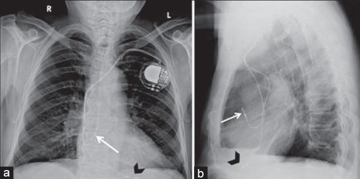
How the implant works
Most pacemakers are placed transvenously: leads are threaded through a vein under the collarbone into the heart, and the generator sits in a small pocket beneath the skin. The device continuously senses the heart's own beats and only fires when the rate drops below its programmed limit — so it works on demand rather than constantly.
The paced ECG
On the ECG, a pacemaker leaves a tell-tale pacing spike — a sharp vertical line — immediately before the wave it triggers. A spike before the QRS means ventricular pacing; a spike before the P wave means atrial pacing. Recognising pacing spikes is a common exam point; practise on our ECG strip questions.
Living with a pacemaker
Modern devices last many years and are checked remotely or in clinic. Most household electronics are safe; strong magnetic fields are the main caution, and many newer pacemakers are MRI-conditional. Patients carry a device ID card.
Key takeaways
- A pacemaker paces the heart when its own rhythm is too slow.
- Main indications: symptomatic bradycardia, AV block, and sick sinus syndrome.
- Types: single-chamber, dual-chamber, biventricular (CRT), and leadless.
- Leads are usually placed transvenously with the generator under the skin.
- A pacing spike before the P or QRS is the ECG signature.
Practise paced-rhythm recognition
Spot pacing spikes and bradyarrhythmias with instant feedback.
Practise ECG Strips →Frequently asked questions
What does a pacemaker do?
It delivers small electrical impulses to make the heart beat when its natural rhythm is too slow, working on demand only when the rate drops below a set limit.
Why would someone need a pacemaker?
For symptomatic bradycardia, high-grade or complete AV block, or sick sinus syndrome — conditions where the heart beats too slowly or unreliably.
What are the types of pacemaker?
Single-chamber, dual-chamber, biventricular (cardiac resynchronization therapy), and leadless pacemakers.
How is a pacemaker implanted?
Usually transvenously: leads are threaded through a vein under the collarbone into the heart and the generator is placed in a pocket beneath the skin.
What does a paced beat look like on an ECG?
A sharp pacing spike appears immediately before the wave it triggers — before the QRS for ventricular pacing or before the P wave for atrial pacing.
Can you have an MRI with a pacemaker?
Many modern pacemakers are MRI-conditional and allow scanning under specific protocols, but older devices may not; it must be checked first.
Sources & further reading
- Cardiovascular Credentialing International (CCI)
- American College of Cardiology
- American Heart Association
- MedlinePlus (U.S. National Library of Medicine)
External links are provided for reference; always confirm current details with the official source.