RCIS Cardiac Anatomy Study Guide
The cardiac anatomy foundations the RCIS exam builds on — chambers, valves, coronaries, and conduction.
Quick facts
- 4 chambers: right atrium, right ventricle, left atrium, left ventricle.
- 4 valves: tricuspid & mitral (AV valves), pulmonic & aortic (semilunar valves).
- 3 major coronaries: LAD, circumflex (from the left main), and RCA.
- Dominance is defined by which artery gives the PDA — RCA in ~85% (right dominant).
- Conduction: SA node → AV node → bundle of His → bundle branches → Purkinje fibers.
What you will learn
- Name the chambers, walls, and septa of the heart.
- Match each valve to its location and leaflet count.
- Trace blood through the systemic and pulmonary circuits.
- Map each coronary artery to the wall it supplies.
- Follow an impulse through the conduction system.
Chambers & walls
The heart has four chambers. The two atria receive blood; the two ventricles pump it. The interatrial septum separates the atria (its remnant of the fetal foramen ovale is the fossa ovalis), and the thicker interventricular septum separates the ventricles.
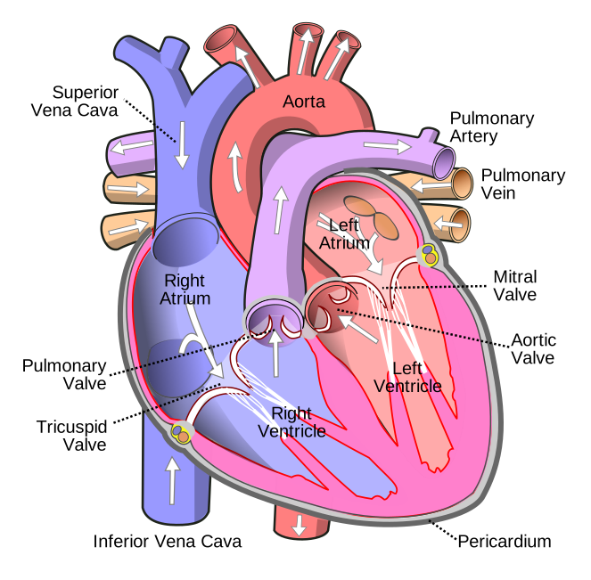
| Chamber | Receives from | Pumps to |
|---|---|---|
| Right atrium | SVC, IVC, coronary sinus | Right ventricle |
| Right ventricle | Right atrium | Pulmonary artery → lungs |
| Left atrium | Four pulmonary veins | Left ventricle |
| Left ventricle | Left atrium | Aorta → body (thickest wall) |
The four heart valves
| Valve | Between | Type | Leaflets |
|---|---|---|---|
| Tricuspid | RA → RV | Atrioventricular | 3 |
| Pulmonic | RV → PA | Semilunar | 3 |
| Mitral (bicuspid) | LA → LV | Atrioventricular | 2 |
| Aortic | LV → aorta | Semilunar | 3 |
Blood flow through the heart
Follow one drop of blood:
Body → SVC/IVC → right atrium → tricuspid → right ventricle → pulmonic valve → pulmonary artery → lungs (picks up oxygen) → pulmonary veins → left atrium → mitral valve → left ventricle → aortic valve → aorta → body.
The right heart is a low-pressure pulmonary pump; the left heart is a high-pressure systemic pump — which is why the left ventricle has the thickest wall.
Coronary arteries & the wall each supplies
The left main coronary artery divides into the LAD and circumflex; the RCA arises separately. Mapping artery → wall is exactly how ECG lead groups localise an MI.
| Artery | Supplies | MI shows in |
|---|---|---|
| LAD (left anterior descending) | Anterior wall, septum, apex | V1–V4 |
| Circumflex | Lateral wall | I, aVL, V5–V6 |
| RCA (right coronary) | Inferior wall, SA/AV nodes (usually) | II, III, aVF |
Coronary dominance
Dominance is defined by which artery gives rise to the posterior descending artery (PDA):
- Right dominant (~85%) — PDA from the RCA.
- Left dominant (~8%) — PDA from the circumflex.
- Co-dominant (~7%) — contributions from both.
The conduction system
The electrical impulse follows a fixed path:
SA node → AV node → bundle of His → right & left bundle branches → Purkinje fibers.
- SA node — the natural pacemaker, in the upper right atrium near the SVC (~60–100 bpm).
- AV node — delays the impulse so the atria empty before the ventricles fire (~40–60 bpm escape).
- Purkinje / ventricular — slowest backup pacemaker (~20–40 bpm).
Exam tips
- Mitral valve = 2 leaflets; the other three valves = 3.
- Coronaries arise from the aortic sinuses of Valsalva.
- Learn artery → wall → ECG leads together; they are one concept.
- Dominance = who gives the PDA.
- SA → AV → His → bundle branches → Purkinje, with descending intrinsic rates.
Summary & key takeaways
- Four chambers, four valves, two circuits — know the flow path cold.
- LAD/circumflex/RCA map to anterior/lateral/inferior walls.
- Dominance is defined by the origin of the PDA.
- The conduction system runs SA → AV → His → bundles → Purkinje.
Frequently asked questions
Where do the coronary arteries originate?
From the aortic sinuses (sinuses of Valsalva) just above the aortic valve.
What is coronary dominance?
It is defined by which artery gives rise to the PDA — the RCA in right dominance, the circumflex in left dominance.
How many chambers and valves does the heart have?
Four chambers (two atria, two ventricles) and four valves (tricuspid, pulmonic, mitral, aortic).
Which heart valve has only two leaflets?
The mitral (bicuspid) valve; the tricuspid, pulmonic, and aortic valves each have three.
Which coronary artery supplies the inferior wall?
The right coronary artery in most people, which is why an inferior MI appears in leads II, III, and aVF.
What is the path of the cardiac conduction system?
SA node → AV node → bundle of His → right and left bundle branches → Purkinje fibers.
Sources & further reading
- Cardiovascular Credentialing International (CCI)
- American College of Cardiology
- American Heart Association
- MedlinePlus (U.S. National Library of Medicine)
External links are provided for reference; always confirm current details with the official source.