RCIS ECG Study Guide
The ECG essentials the RCIS exam tests — waves, intervals, and the arrhythmias you must recognise.
Quick facts
- P wave = atrial depolarization · QRS = ventricular depolarization · T wave = ventricular repolarization.
- Normal intervals: PR 0.12–0.20 s · QRS < 0.12 s · QTc < 0.44 s.
- Paper speed 25 mm/s: one small box = 0.04 s, one large box = 0.20 s. 10 mm = 1 mV.
- Rate shortcut: 300 ÷ (large boxes between R waves) → 300, 150, 100, 75, 60, 50.
- Emergencies: VFib / pulseless VT → defibrillate; asystole → CPR + epinephrine (never shock).
What you will learn
- Read any strip with a repeatable 5-step method.
- Calculate heart rate three ways.
- Recall normal intervals and what widens or prolongs them.
- Recognise the core arrhythmias on sight.
- Distinguish the AV blocks and know the emergency rhythms cold.
A systematic 5-step approach
Read every strip the same way so nothing is missed:
- Rate — fast, slow, or normal?
- Rhythm — regular or irregular? Pattern?
- P waves — present, and one before every QRS?
- PR interval — normal, long, or variable?
- QRS — narrow (supraventricular) or wide (ventricular / bundle branch block)?
Waves, segments, and intervals
| Component | Represents | Normal |
|---|---|---|
| P wave | Atrial depolarization | upright in lead II |
| PR interval | Atrial depol + AV delay | 0.12–0.20 s |
| QRS complex | Ventricular depolarization | < 0.12 s |
| ST segment | Early repolarization | isoelectric |
| T wave | Ventricular repolarization | upright in lead II |
| QT interval | Total ventricular activity | QTc < 0.44 s |
Calculating heart rate
- 300 method (regular): 300 ÷ number of large boxes between two R waves.
- 1500 method (precise): 1500 ÷ number of small boxes between two R waves.
- 6-second method (irregular): count QRS complexes in a 6-second strip and multiply by 10.
Under 60 bpm is bradycardia; over 100 bpm is tachycardia.
Core rhythms to recognise on sight
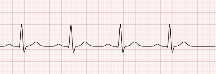
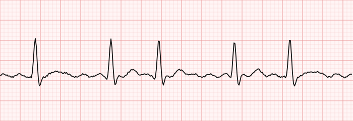
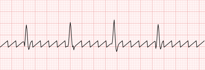
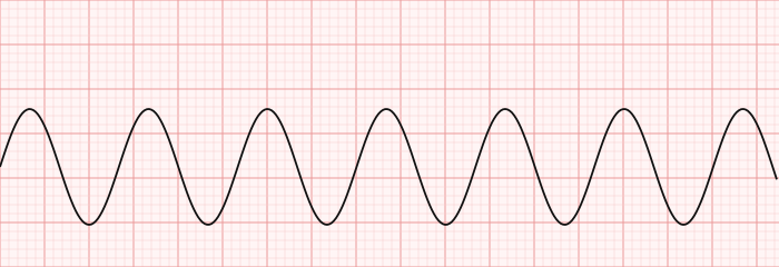
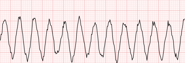
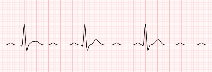
Test yourself on real strips in our ECG strip identification questions.
AV blocks
| Block | Hallmark |
|---|---|
| First-degree | PR > 0.20 s, every P conducts |
| Mobitz I (Wenckebach) | PR lengthens progressively, then a beat drops |
| Mobitz II | Constant PR, sudden dropped beat (can progress to complete block) |
| Third-degree (complete) | P waves and QRS march independently |
Emergency rhythms & STEMI localization
- VFib / pulseless VT — defibrillate immediately + CPR.
- Asystole / PEA — CPR + epinephrine; not shockable.
- Symptomatic bradycardia — atropine, then pacing.
Localise an ST-elevation MI by lead group:
| Leads | Wall | Usual artery |
|---|---|---|
| II, III, aVF | Inferior | RCA |
| V1–V4 | Anteroseptal | LAD |
| I, aVL, V5–V6 | Lateral | Circumflex |
Exam tips
- Always work the 5 steps in order — rate, rhythm, P, PR, QRS.
- Irregularly irregular with no P waves = atrial fibrillation.
- Sawtooth = flutter; chaotic + pulseless = VFib (shock it).
- Wide + regular + fast = VT until proven otherwise.
- Tall, tented T waves = hyperkalemia.
Summary & key takeaways
- Use one systematic method for every strip.
- Know normal intervals and the three rate-calculation methods.
- Recognise AFib, flutter, VT, VFib, and complete block instantly.
- Match the AV blocks to their hallmark pattern.
- Memorise the shockable rhythms and the STEMI lead groups.
Frequently asked questions
What does the QRS complex represent?
Ventricular depolarization; a normal QRS is under 0.12 seconds.
How is ventricular fibrillation treated?
Immediate defibrillation with CPR — it is a pulseless emergency.
What is the normal PR interval?
0.12 to 0.20 seconds (3 to 5 small boxes). A PR over 0.20 s is first-degree AV block.
How do you calculate heart rate on an ECG?
Divide 300 by the number of large boxes between two R waves for a regular rhythm, or count QRS complexes in a 6-second strip and multiply by 10 for an irregular one.
How do you tell VT from SVT?
Ventricular tachycardia is a wide-complex (≥0.12 s) regular tachycardia; SVT is narrow-complex. Treat a wide regular tachycardia as VT until proven otherwise.
Which ECG leads show an inferior MI?
Leads II, III, and aVF, usually from a right coronary artery occlusion.
Sources & further reading
- Cardiovascular Credentialing International (CCI)
- American College of Cardiology
- American Heart Association
- MedlinePlus (U.S. National Library of Medicine)
External links are provided for reference; always confirm current details with the official source.