Coronary Artery Disease (CAD): Causes, Symptoms & Treatment
Coronary artery disease is the slow narrowing of the heart’s own arteries by fatty plaque — the single most common cause of death worldwide, and the quiet setup behind most heart attacks. Understanding how it starts, how it shows up, and how it is treated is foundational for anyone working in cardiovascular care.
What is coronary artery disease?
Coronary artery disease (CAD) — sometimes called coronary heart disease or ischemic heart disease — is the buildup of fatty, cholesterol-rich plaque inside the arteries that supply blood to the heart muscle. As these plaques grow, they narrow the vessel and stiffen its walls, choking the flow of oxygen-rich blood the heart needs to keep pumping. When demand outstrips supply, the muscle becomes ischemic, and the person feels it as chest discomfort or breathlessness.
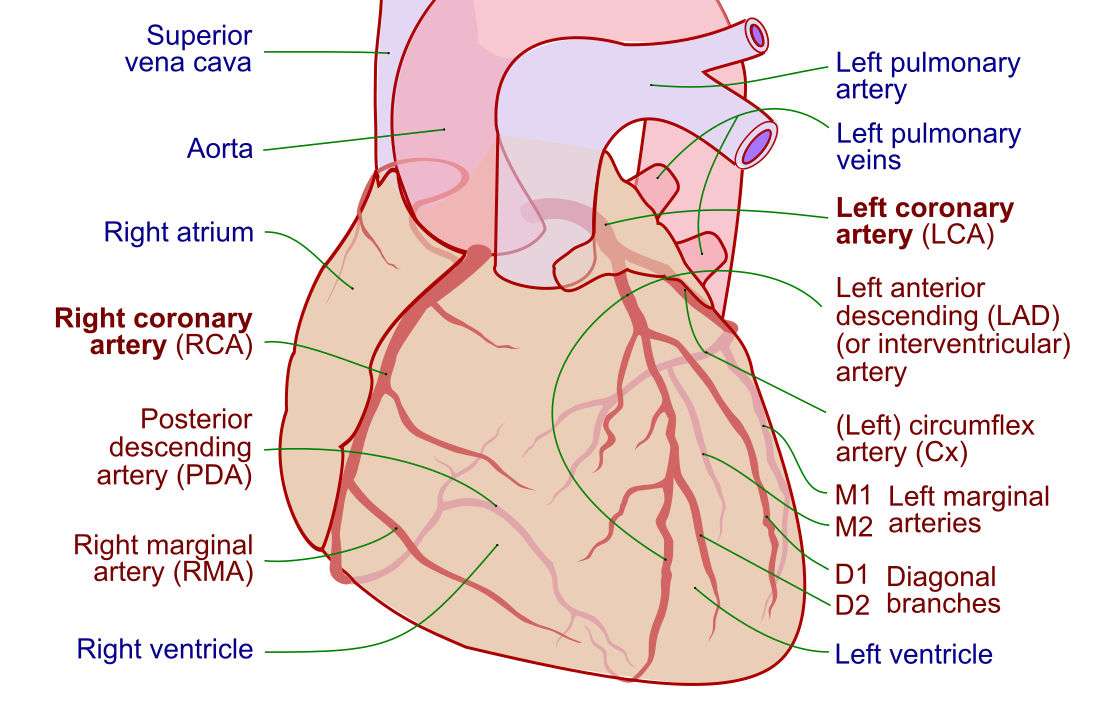
The heart is a tireless pump, but it cannot draw oxygen from the blood rushing through its own chambers. It relies on a dedicated network of vessels — the coronary arteries — that arch across its surface and dive into the muscle. A working knowledge of coronary artery anatomy makes CAD far easier to reason about, because which artery narrows determines which part of the heart suffers.
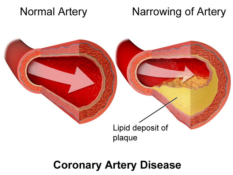
CAD exists on a spectrum. At one end sits chronic (stable) coronary disease, where narrowing is fixed and symptoms are predictable. At the other end lie the acute coronary syndromes — unstable angina, NSTEMI, and STEMI — where a plaque suddenly ruptures and a clot forms, turning a manageable narrowing into an emergency. The vast majority of heart attacks are simply CAD that has crossed that line.
This article is written for students and clinicians preparing for exams such as the RCIS. It is educational and not a substitute for medical advice; anyone with chest pain or suspected heart problems should be evaluated by a clinician.
Atherosclerosis: the root cause
The engine driving CAD is atherosclerosis — a decades-long inflammatory process in the artery wall. It rarely announces itself. Plaque can accumulate silently from young adulthood, and many people have significant disease long before a single symptom appears.
The story usually begins with injury to the delicate inner lining of the artery, the endothelium. High blood pressure, tobacco smoke, elevated LDL cholesterol, and high blood sugar all batter this lining. Once it is damaged, LDL particles slip beneath the surface, become oxidized, and trigger an immune response. White blood cells move in, engulf the fat, and turn into foam cells, forming an early fatty streak. Over years, this evolves into a mature plaque with a lipid-rich core capped by fibrous tissue.
Not all plaques behave the same way, and this distinction is central to how CAD kills:
- Stable plaques have a thick, tough fibrous cap. They narrow the artery gradually and tend to produce predictable, exertional symptoms.
- Vulnerable plaques have a thin, inflamed cap over a soft, fatty core. They may not narrow the vessel dramatically — but they are prone to rupture. When the cap tears, the body treats it as a wound, platelets swarm the exposed core, and a clot forms that can occlude the artery in minutes.
This is the crucial insight for exams and for practice: the plaque that causes a heart attack is often not the biggest one. A modestly narrowed but unstable lesion can rupture and kill, while a tightly narrowed but stable lesion may only cause angina. That mismatch explains why a person can pass a stress test and still have a heart attack weeks later.
Risk factors you can and cannot change
CAD is a disease of accumulated risk. No single factor causes it; instead, several forces compound over time to accelerate atherosclerosis. Guidelines split these into factors you cannot modify and — more usefully — those you can.
| Non-modifiable | Modifiable |
|---|---|
| Age (risk rises with each decade) | Smoking and tobacco use |
| Male sex / postmenopausal status | High LDL cholesterol |
| Family history of early heart disease | Hypertension |
| Genetic conditions (e.g., familial hypercholesterolemia) | Diabetes and insulin resistance |
| Ethnic background | Obesity, physical inactivity, poor diet |
The modifiable list is where prevention lives. Current guidance emphasizes estimating a person’s overall 10-year cardiovascular risk rather than fixating on any single number, then treating the whole picture. Smoking cessation, LDL lowering with statins, blood pressure control, and management of diabetes each independently reduce events — and their benefits stack.
Several so-called risk enhancers have gained attention as the evidence evolves: chronic inflammatory conditions, chronic kidney disease, a high lipoprotein(a), metabolic syndrome, and adverse pregnancy history can all nudge risk upward and tip borderline patients toward treatment. This is an area where recommendations continue to be refined, so the emphasis is on individualized, shared decision-making rather than rigid thresholds.
Angina and other symptoms
The classic symptom of CAD is angina — chest discomfort caused by the heart muscle not getting enough oxygen. Patients rarely describe it as sharp pain. More often it is a pressure, tightness, squeezing, or heaviness behind the breastbone, sometimes radiating to the left arm, jaw, neck, or back. It is the heart’s way of signaling that supply has fallen short of demand.
The pattern of angina matters as much as its presence, because it separates stable disease from an emergency:
- Stable angina is predictable. It comes on with exertion or emotional stress, follows a consistent threshold, and eases within minutes of rest or nitroglycerin. It reflects a fixed narrowing that cannot supply enough blood when the heart works harder.
- Unstable angina is a warning of impending catastrophe. It occurs at rest, is new, or is clearly worsening in frequency or intensity. It signals a plaque that has become unstable and belongs to the acute coronary syndromes — a call-emergency-services situation.
Beyond chest discomfort, CAD can present with shortness of breath, unusual fatigue, lightheadedness, nausea, or a cold sweat. Crucially, some people — particularly women, older adults, and those with diabetes — have atypical or silent presentations, with breathlessness or fatigue instead of chest pain, or no symptoms until a heart attack strikes.
When ischemia does progress to muscle death, the presentation becomes a myocardial infarction, and the priorities shift entirely from evaluation to rapid reperfusion.
How CAD is diagnosed
Diagnosing CAD blends the clinical story with tests that either reveal ischemia (the functional consequence) or visualize the plaque itself (the anatomy). No single test is perfect; they are chosen based on a patient’s pretest likelihood of disease.
The workup usually starts simply. A resting 12-lead ECG may be normal in stable disease but can show old infarcts or ischemic changes; solid grounding in ECG rhythm interpretation helps catch the arrhythmias that CAD can trigger. Blood work assesses cholesterol, glucose, and — when an acute event is suspected — cardiac troponin.
From there, testing branches into two families:
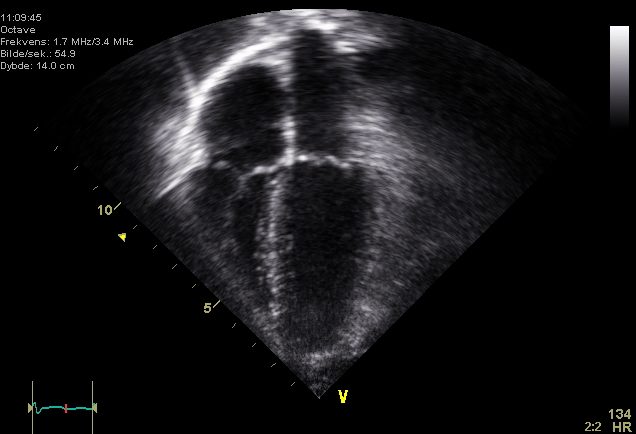
- Functional tests look for ischemia when the heart is stressed. A cardiac stress test — with exercise or pharmacologic stress — can be paired with ECG, an echocardiogram, or nuclear imaging to see whether a region of muscle is starved under load.
- Anatomic tests visualize the arteries directly. Coronary CT angiography (CCTA) has become a first-line non-invasive way to rule out significant disease, while invasive coronary angiography in the cath lab remains the gold standard — and can move seamlessly into treatment.
In the cath lab, angiography does more than picture the plaque. Physiologic measurements such as fractional flow reserve gauge whether a given narrowing is actually limiting flow, and a good grasp of hemodynamic principles underpins how these decisions are made. The result is a shift from treating every narrowing to treating only those that matter functionally.
Medical treatment and lifestyle
For most people with stable CAD, the foundation of treatment is not a procedure — it is guideline-directed medical therapy paired with lifestyle change. Landmark trials have shown that, in stable disease, optimal medications control symptoms and reduce events comparably to routine stenting in many patients, reserving revascularization for those who fail medical therapy or have high-risk anatomy.
The medication strategy targets two goals: relieving symptoms and preventing future events.
- Antiplatelet therapy — low-dose aspirin is a mainstay for those with established disease, keeping platelets from clumping on plaque.
- High-intensity statins — these lower LDL and, just as importantly, stabilize plaque and calm inflammation. Additional LDL-lowering agents are added when targets are not met.
- Anti-anginal drugs — beta-blockers, calcium channel blockers, and nitroglycerin ease the heart’s workload and dilate vessels to relieve angina.
- Blood pressure and diabetes control — agents such as ACE inhibitors provide added protection, particularly in patients with reduced heart function or diabetes.
Medications only go so far without lifestyle change, which is genuinely disease-modifying. Stopping smoking, adopting a heart-healthy diet, exercising regularly, and managing weight and stress all slow atherosclerosis and cut event rates. These are not soft recommendations — smoking cessation alone produces one of the largest single reductions in cardiovascular risk available.
Revascularization: PCI vs CABG
When narrowing is severe, symptoms persist despite medication, or a patient presents with an acute coronary syndrome, the goal becomes revascularization — restoring blood flow past the blockage. Two strategies dominate: percutaneous coronary intervention and coronary artery bypass grafting.
Percutaneous coronary intervention (PCI) is the catheter-based approach most people call angioplasty with a stent. A thin catheter is threaded from the wrist or groin to the coronary artery, a balloon is inflated to crack open the narrowing, and a drug-eluting stent is deployed to scaffold the vessel open. It is minimally invasive, done under local anesthesia, and is the default emergency treatment for a STEMI. Our comparison of PCI versus diagnostic catheterization and an overview of what happens in a cardiac cath lab flesh out the procedure.
Coronary artery bypass grafting (CABG) is open-heart surgery. A surgeon harvests a healthy vessel — typically the internal mammary artery or a leg vein — and sews it in to route blood around the blockage. It is more invasive, with a longer recovery, but for the right anatomy it offers durable, complete revascularization.
| Feature | PCI (stent) | CABG (surgery) |
|---|---|---|
| Approach | Catheter through wrist/groin | Open-heart surgery |
| Anesthesia | Local + sedation | General |
| Recovery | Days | Weeks |
| Best suited for | One or two vessels; acute STEMI | Left main or complex multi-vessel disease; diabetes |
| Durability | Good; some restenosis risk | Excellent, especially arterial grafts |
Choosing between them is a team decision. In general, PCI shines for focal disease and emergencies, while CABG tends to win for left main disease, extensive multi-vessel disease, and patients with diabetes, where trials show a survival or durability advantage. Anatomic complexity scores and the patient’s overall health guide this “heart team” discussion — and, as evidence accumulates, the boundaries keep shifting.
Complications and long-term outlook
Left unchecked, CAD progresses, and its complications are exactly why it remains the leading cause of death worldwide. The most feared is the heart attack — a ruptured plaque and clot that abruptly kill muscle. But there are others clinicians watch for.
Chronic ischemia and prior infarcts weaken the pump. When enough muscle is damaged or hibernating, the heart’s output falls and ischemic cardiomyopathy or heart failure sets in; a falling ejection fraction often marks this transition and shapes prognosis. Scarred, irritable muscle is also electrically unstable, raising the risk of dangerous rhythms such as ventricular tachycardia and sudden cardiac death.
In severe acute events, a large infarct can drop cardiac output so far that organs are underperfused — cardiogenic shock. Managing these patients draws on a deep understanding of shock hemodynamics, and the sickest may need temporary mechanical support such as an intra-aortic balloon pump while the heart recovers.
The outlook, though, has improved dramatically. With modern statins, antiplatelets, timely revascularization, and aggressive risk-factor control, many people with CAD live long, active lives. The emphasis has shifted from crisis management to prevention: slowing atherosclerosis, stabilizing plaque, and stopping the first — or next — heart attack before it happens. Cardiac rehabilitation, combining supervised exercise with education and risk-factor management, measurably improves survival and quality of life after an event.
Key takeaways
- Coronary artery disease is the narrowing of the heart’s arteries by plaque, driven by atherosclerosis, and is the world’s leading cause of death.
- The plaque that triggers a heart attack is often not the largest but the most vulnerable — a thin-capped, inflamed lesion prone to rupture and clot.
- Angina is the hallmark symptom; stable angina is predictable and exertional, while unstable angina occurs at rest and signals an acute coronary syndrome.
- Diagnosis combines the clinical story with functional tests (stress testing, echo) and anatomic imaging (CT angiography, invasive angiography).
- In stable disease, medications and lifestyle come first; statins, aspirin, anti-anginals, and smoking cessation are foundational.
- PCI versus CABG: PCI (stenting) suits focal disease and emergencies; CABG suits left main, complex multi-vessel disease, and many patients with diabetes.
- Complications include heart attack, heart failure, dangerous arrhythmias, and cardiogenic shock — but prevention and rehab dramatically improve outcomes.
- This content is educational, not medical advice; anyone with chest pain or suspected CAD should seek prompt medical evaluation.
Learn how CAD is treated in the lab
Understand PCI, stenting, and cardiac catheterization.
PCI vs Cardiac Cath →Frequently asked questions
What is coronary artery disease in simple terms?
Coronary artery disease is the buildup of fatty plaque inside the arteries that supply blood to the heart muscle. As the plaque grows, it narrows the arteries and reduces the flow of oxygen-rich blood, which can cause chest discomfort (angina) and, if a plaque ruptures and a clot forms, a heart attack. It is a slow, often silent process that develops over many years.
What is the difference between coronary artery disease and a heart attack?
Coronary artery disease is the underlying condition — the gradual narrowing of the coronary arteries by plaque. A heart attack (myocardial infarction) is an acute event that happens when a plaque ruptures and a clot suddenly blocks an artery, killing heart muscle. In short, CAD is the chronic disease, and a heart attack is the emergency it can cause.
What are the warning signs of coronary artery disease?
The most common sign is angina — chest pressure, tightness, or squeezing, often brought on by exertion and radiating to the arm, jaw, neck, or back. Other signs include shortness of breath, unusual fatigue, lightheadedness, nausea, or a cold sweat. Some people, especially women, older adults, and those with diabetes, have atypical symptoms or none at all until a heart attack occurs.
Can coronary artery disease be reversed?
CAD generally cannot be fully reversed, but its progression can be slowed, halted, or even modestly improved. Aggressive LDL lowering with statins, smoking cessation, a heart-healthy diet, regular exercise, and control of blood pressure and diabetes can stabilize plaque and reduce the risk of heart attacks. The realistic goal is to stop the disease from advancing and to prevent events.
What is the difference between PCI and CABG?
PCI (percutaneous coronary intervention) is a minimally invasive procedure that uses a catheter to open a narrowed artery and place a stent. CABG (coronary artery bypass grafting) is open-heart surgery that routes blood around blockages using grafted vessels. PCI is preferred for focal disease and emergencies like STEMI, while CABG is often better for left main disease, complex multi-vessel disease, and many patients with diabetes.
How is coronary artery disease diagnosed?
Diagnosis combines the patient’s symptoms and risk factors with testing. Functional tests such as a stress test with ECG, echocardiogram, or nuclear imaging look for ischemia, while anatomic tests such as coronary CT angiography or invasive coronary angiography visualize the arteries directly. Blood tests for cholesterol and, when an acute event is suspected, cardiac troponin round out the workup.
What causes coronary artery disease?
CAD is caused by atherosclerosis — a slow, inflammatory buildup of cholesterol-rich plaque in the artery walls. Key contributing factors include smoking, high LDL cholesterol, high blood pressure, diabetes, obesity, physical inactivity, and a family history of early heart disease. These factors damage the artery lining and accelerate plaque formation over many years.
Is angina the same as a heart attack?
No. Angina is chest discomfort from reduced blood flow that does not kill heart muscle — it is a warning sign. Stable angina comes on with exertion and eases with rest. A heart attack involves prolonged blockage that actually damages heart muscle and is confirmed by a rise in troponin. Unstable angina, which occurs at rest or worsens, sits in between and is treated as an emergency.
Can you live a normal life with coronary artery disease?
Yes. Many people with CAD live long, active lives, especially with modern treatment. Statins, antiplatelet therapy, blood pressure and diabetes control, timely revascularization when needed, and cardiac rehabilitation all improve outcomes. Consistent lifestyle changes — not smoking, eating well, and staying active — are central to living well with the condition.
Sources & further reading
- Cardiovascular Credentialing International (CCI)
- American College of Cardiology
- American Heart Association
- MedlinePlus (U.S. National Library of Medicine)
External links are provided for reference; always confirm current details with the official source.