Cardiac Stress Test: Types, What to Expect & Results
A cardiac stress test pushes the heart harder than rest ever asks it to — and watches for the electrical, imaging, or symptom clues that quietly signal a coronary artery struggling to keep up. This guide breaks down how the exercise and pharmacologic versions work, what nuclear and echo add, and how to read the results.
- What is a cardiac stress test?
- Why and when stress testing is ordered
- Exercise vs pharmacologic stress: choosing the trigger
- Pharmacologic agents: Lexiscan, adenosine, and dobutamine
- Nuclear stress vs stress echo: the imaging layer
- What happens during the test
- Reading the results: what positive and negative mean
- Limitations, risks, and next steps
- Key takeaways
What is a cardiac stress test?
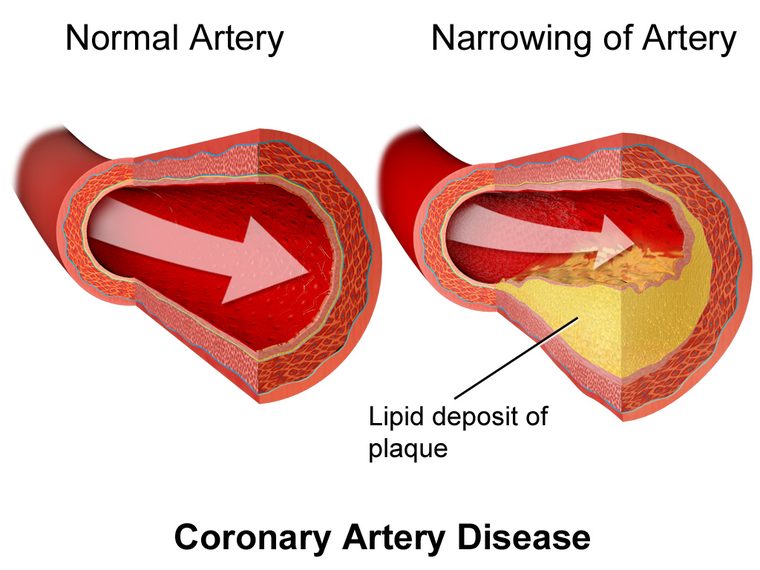
A cardiac stress test is a controlled way of asking the heart to work harder while clinicians monitor how it responds. At rest, a narrowed coronary artery can still deliver enough blood to keep the heart muscle happy, so a resting ECG or a calm patient may look completely normal. The disease hides. Stress testing unmasks it by ramping up the heart's demand for oxygen — through exercise or medication — and watching for the mismatch between what the muscle needs and what the arteries can supply.
That mismatch, called ischemia, shows up in predictable ways: ST-segment shifts on the ECG, new wall-motion abnormalities on ultrasound, reduced tracer uptake on nuclear imaging, a drop in blood pressure, or classic anginal symptoms. The test doesn't image the plaque directly the way a CT angiogram or a cath does; instead it measures the functional consequence of a blockage. That distinction matters, because a coronary lesion that looks impressive on an angiogram may or may not actually limit flow, and stress testing helps sort out which lesions are doing real harm.
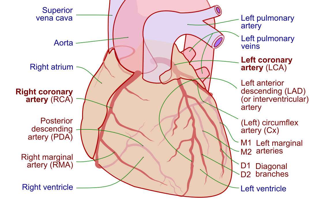
Stress testing sits at the crossroads of cardiology, and understanding it rewards a solid grasp of underlying coronary artery anatomy and the way coronary artery disease progresses from a soft plaque to a flow-limiting stenosis. This article is educational and written for students and clinicians building foundational knowledge; it is not medical advice, and no one should interpret their own test results without their care team.
Why and when stress testing is ordered
The most common reason to order a stress test is stable chest pain in a patient with an intermediate likelihood of coronary disease. If the pretest probability is very low, a positive result is more likely to be a false alarm than real disease; if it is very high, the patient may go straight to the cath lab. Stress testing earns its keep in the broad middle, where the result genuinely changes management.
Beyond diagnosis, stress tests are used to:
- Risk-stratify known coronary disease — how much myocardium is at risk, and how severe is the ischemia?
- Assess functional capacity before major noncardiac surgery or as part of cardiac rehab planning.
- Evaluate the effect of therapy — did medication or a stent actually improve the patient's ischemic burden?
- Provoke arrhythmias or exercise-induced symptoms that don't appear at rest.
Current guideline thinking has shifted somewhat toward CT coronary angiography as a first-line test in many stable chest-pain patients, especially younger ones without prior disease, because it directly visualizes anatomy. Stress testing remains firmly in the toolkit, though — particularly when a functional answer is what clinicians actually need, when radiation or contrast must be minimized, or when the patient already has known disease and the question is about ischemia, not the presence of plaque. The best test is the one matched to the specific clinical question, and that judgment is still evolving as imaging technology improves.
Exercise vs pharmacologic stress: choosing the trigger
Every stress test needs a stressor. The two broad families are exercise and pharmacologic, and the choice hinges mostly on whether the patient can walk hard enough to reach a diagnostic level of exertion.
Exercise stress is the preferred approach whenever feasible. Walking on a treadmill (usually the Bruce protocol, with its stepwise increases in speed and incline) or pedaling a bike is physiologic — it raises heart rate and blood pressure the way real life does, and it yields bonus information you can't get from a drug: how long the patient lasted, their peak workload in METs, their blood-pressure response, their symptoms, and their heart-rate recovery afterward. Exercise capacity itself is one of the strongest predictors of long-term outcome, sometimes more telling than the ischemia findings.
The goal in an exercise test is to push the patient to at least 85% of age-predicted maximum heart rate (a rough estimate of that maximum is 220 minus age). Fall short of that target and a negative test becomes hard to trust, because the heart may simply not have been challenged enough to reveal a limiting lesion.
Pharmacologic stress steps in when a patient can't exercise adequately — because of arthritis, peripheral arterial disease, deconditioning, amputation, severe lung disease, or an inability to follow the protocol. Instead of physical exertion, a drug creates the stress. The catch is that pharmacologic stress usually must be paired with an imaging technique (nuclear or echo), because these drugs don't reliably produce the ECG changes that a plain treadmill test relies on.
| Feature | Exercise stress | Pharmacologic stress |
|---|---|---|
| Trigger | Treadmill or bike | Vasodilator or dobutamine |
| Preferred when | Patient can exercise | Patient cannot exercise adequately |
| Bonus data | METs, BP response, HR recovery, symptoms | Limited functional data |
| Usual pairing | Can be ECG-only or with imaging | Almost always with imaging |
| Diagnostic ECG value | High | Low — imaging carries the diagnosis |
Pharmacologic agents: Lexiscan, adenosine, and dobutamine
Pharmacologic agents split into two mechanistic camps, and knowing the difference is a favorite exam target as well as a genuine clinical decision point.
Vasodilators: regadenoson (Lexiscan), adenosine, dipyridamole
Regadenoson — brand name Lexiscan — is now the most widely used vasodilator for stress imaging in the United States. Along with adenosine and dipyridamole, it works by dilating the coronary microvasculature. Here's the elegant trick: normal arteries dilate fully and their flow surges, while a stenosed artery is already maximally dilated at rest and can't increase flow much further. The result is heterogeneous flow — healthy territory lights up with tracer or contrast, diseased territory lags behind. That flow disparity, not true ischemia, is what nuclear and (increasingly) echo imaging detect.
Lexiscan's popularity comes from convenience: it's a fixed single-dose bolus, its A2A-receptor selectivity means fewer bronchospastic side effects than older adenosine, and it doesn't require a weight-based infusion. Common transient side effects include flushing, headache, chest tightness, and shortness of breath. Because these agents can provoke bronchospasm and high-grade AV block, patients with severe reactive airway disease or significant conduction disease need careful selection, and aminophylline is kept on hand as a reversal agent. Patients are typically asked to avoid caffeine and other methylxanthines for 12 or more hours beforehand, since these compounds block the adenosine receptors the drugs act on and can blunt the test.
Inotrope/chronotrope: dobutamine
Dobutamine takes a different route. Rather than dilating vessels, it directly stimulates the heart — increasing contractility, heart rate, and blood pressure to mimic the demand of exercise. It's the go-to agent when vasodilators are contraindicated, classically in patients with bronchospastic lung disease. Dobutamine is infused in escalating doses, and atropine is often added to help reach the target heart rate. Because it drives the heart so hard, dobutamine can provoke arrhythmias, and the infusion is titrated carefully with continuous monitoring.
- Lexiscan / adenosine / dipyridamole = dilate → flow heterogeneity (a supply test)
- Dobutamine = drive → increased demand, exercise-mimicking (a demand test)
Whichever agent is chosen, the pharmacology overlaps with the broader world of cath lab medications, and clinicians managing these patients should be fluent in both the drug effects and their antidotes.
Nuclear stress vs stress echo: the imaging layer
When a stress test needs imaging — always with pharmacologic stress, and often with exercise too — the two dominant modalities are nuclear perfusion imaging and stress echocardiography. They answer the same clinical question through very different physics.
Nuclear stress (SPECT / PET myocardial perfusion imaging)
In nuclear imaging, a small amount of radioactive tracer (technetium-based agents, or rubidium/ammonia for PET) is injected into the bloodstream. The tracer is taken up by heart muscle in proportion to blood flow. A gamma camera or PET scanner then maps where the tracer went. Images are captured both at stress and at rest, and the two are compared:
- A reversible defect — abnormal at stress, normal at rest — signals ischemia from a flow-limiting stenosis.
- A fixed defect — abnormal on both — usually indicates scar from a prior myocardial infarction.
Nuclear imaging is highly sensitive and quantifiable, and PET adds the ability to measure absolute myocardial blood flow. The trade-offs are radiation exposure, higher cost, and the logistics of a radiopharmaceutical.
Stress echocardiography
Stress echo uses ultrasound instead of radiation. The sonographer captures images of the heart's walls at rest and again at peak stress, and the reader compares wall motion. Normally, every segment of the left ventricle thickens and moves inward more vigorously under stress. A segment that fails to augment — or actually moves worse — betrays ischemia in that coronary territory. Stress echo is radiation-free, portable, and cheaper, and it delivers a wealth of extra structural data (valves, ejection fraction, chamber sizes). Its main limitations are operator dependence and image quality in patients with difficult acoustic windows; contrast agents help in tough cases. A strong grounding in standard echocardiography is essential before tackling the stress version.
| Nuclear (SPECT/PET) | Stress echo | |
|---|---|---|
| What it shows | Perfusion (tracer uptake) | Wall motion |
| Radiation | Yes | None |
| Extra anatomy | Limited | Valves, EF, chambers |
| Operator dependence | Lower | Higher |
| Cost | Higher | Lower |
What happens during the test
Whatever flavor is ordered, the day follows a recognizable arc. Patients are usually asked to arrive fasting, to hold certain medications (beta-blockers and calcium channel blockers can blunt an exercise test; caffeine interferes with vasodilators), and to wear comfortable shoes if they'll be walking.
- Prep. Electrodes are placed for continuous ECG monitoring, a blood-pressure cuff is applied, and an IV is started if imaging or drugs are involved. Baseline ECG and vitals are recorded.
- Baseline imaging (if applicable). Rest nuclear images or a resting echo are obtained for comparison.
- Stress. The patient exercises to target, or the drug is infused. Heart rate, rhythm, blood pressure, and symptoms are tracked continuously.
- Peak capture. At maximum stress, tracer is injected or peak echo images are grabbed within the narrow window of highest demand.
- Recovery. Monitoring continues for several minutes. Heart-rate recovery and any delayed ST changes or arrhythmias are noted.
Supervising staff watch for indications to stop early: significant ST-segment depression or elevation, a fall in systolic blood pressure, serious arrhythmias such as ventricular tachycardia, severe symptoms, or the patient simply reaching exhaustion. Safety hinges on rapid rhythm recognition, so practicing ECG strip interpretation is time well spent for anyone staffing these tests.
Reading the results: what positive and negative mean
The headline result of a stress test is usually reported as positive (evidence of ischemia), negative (no evidence), equivocal (uncertain), or non-diagnostic (the test couldn't answer the question — often because the patient didn't reach target heart rate). But the interpretation is richer than a single word.
On the ECG, the classic marker of ischemia is horizontal or downsloping ST-segment depression of at least 1 mm. Upsloping ST depression is far less specific. ST elevation during stress (in leads without prior infarction) is worrying and can signal severe disease — a pattern worth understanding alongside the fundamentals of ST-elevation interpretation. The ECG isn't read in isolation, though: symptoms, blood-pressure response, exercise capacity, and heart-rate recovery all fold into the final read.
On imaging, positivity means a reversible perfusion defect (nuclear) or a stress-induced wall-motion abnormality (echo). The size and location of the abnormality map to specific coronary territories and estimate how much myocardium is jeopardized — a small single-territory defect carries very different weight than a large multi-territory one.
| Finding | Suggests |
|---|---|
| Horizontal/downsloping ST depression ≥1 mm | Ischemia |
| Exercise-induced ST elevation (no prior MI) | Severe/transmural ischemia |
| Fall in systolic BP with exertion | High-risk finding, possible severe CAD |
| Reversible perfusion defect (nuclear) | Flow-limiting stenosis |
| Fixed perfusion defect (nuclear) | Prior infarct / scar |
| New wall-motion abnormality (echo) | Ischemia in that territory |
| Poor exercise capacity, blunted HR recovery | Adverse prognosis independent of ischemia |
Limitations, risks, and next steps
Stress testing is generally very safe, but it isn't risk-free. Because it deliberately provokes cardiac stress, rare serious events — arrhythmia, infarction, or hemodynamic collapse — can occur, which is why testing happens with trained staff and resuscitation equipment on hand. Absolute contraindications include acute MI within the last couple of days, unstable angina not yet stabilized, uncontrolled arrhythmia, symptomatic severe aortic stenosis, and acute conditions like aortic dissection or pulmonary embolism.
The interpretive limits matter just as much as the physical ones. A test that fails to reach target heart rate is not a reassuring negative — it's simply non-diagnostic. Sensitivity and specificity vary by modality, patient sex, body habitus, and baseline ECG. Single-vessel disease and balanced multi-vessel ischemia (where all territories are equally underperfused) can both fool a test into looking normal.
When a stress test is positive or high-risk, the usual next step is invasive coronary angiography in the cath lab, sometimes with revascularization by PCI or bypass. Understanding how findings translate into cath-lab decisions is a core competency for anyone working in interventional cardiology; a tour through the cardiac cath lab puts the whole diagnostic-to-treatment pathway in context. For students preparing credentialing exams, layering stress-test knowledge onto broader hemodynamics fundamentals builds the integrated picture that boards love to test.
Key takeaways
- A cardiac stress test reveals coronary disease that hides at rest by raising the heart's oxygen demand and watching for ischemia.
- Exercise stress is preferred when possible — it adds prognostic data (METs, BP response, heart-rate recovery) a drug can't provide; aim for ≥85% of age-predicted max heart rate.
- Pharmacologic stress is for patients who can't exercise. Vasodilators (Lexiscan/regadenoson, adenosine, dipyridamole) create flow heterogeneity; dobutamine drives demand and is favored in bronchospastic lung disease.
- Hold caffeine before vasodilators and beta-blockers before dobutamine; keep aminophylline and monitoring ready.
- Nuclear imaging maps perfusion (reversible = ischemia, fixed = scar); stress echo maps wall motion, adds no radiation, and shows extra structure.
- Positive results include ≥1 mm horizontal/downsloping ST depression, reversible defects, or new wall-motion abnormalities — always interpreted against pretest probability.
- A test that misses target heart rate is non-diagnostic, not negative.
- This article is educational, not medical advice; guideline preferences between functional and anatomic testing continue to evolve.
Practise cardiovascular fundamentals
Test ischaemia, ECG, and procedure questions.
Practise RCIS Core →Frequently asked questions
How long does a cardiac stress test take?
A basic exercise ECG test often takes about 30 to 60 minutes including prep and recovery, but the actual exercise portion is usually only 7 to 15 minutes. Nuclear stress tests take much longer — often two to four hours — because they require separate rest and stress imaging with waiting time between injections and scans. Stress echo typically falls in between, around an hour.
What is the difference between a nuclear stress test and a stress echo?
A nuclear stress test injects a radioactive tracer and images how well blood (and therefore the tracer) reaches the heart muscle, comparing stress and rest perfusion. A stress echo uses ultrasound to compare how the heart walls move at rest versus peak stress. Nuclear imaging measures perfusion and involves radiation; stress echo measures wall motion, uses no radiation, and shows extra structure like valves and ejection fraction. Both are effective; the choice depends on the patient, local expertise, and the clinical question.
Why do I have to avoid caffeine before a stress test?
Vasodilator agents like Lexiscan (regadenoson), adenosine, and dipyridamole work by acting on adenosine receptors in the coronary microvasculature. Caffeine and related methylxanthines block those same receptors, which can blunt the drug's effect and make the test unreliable or falsely negative. Patients are usually told to avoid caffeine — coffee, tea, soda, chocolate, and some medications — for 12 hours or more beforehand. This restriction doesn't apply to a plain treadmill test the same way, but always follow your specific instructions.
What does a positive stress test mean?
A positive stress test means the study found evidence of ischemia — the heart muscle wasn't getting enough blood under stress. On an ECG that's typically horizontal or downsloping ST-segment depression; on nuclear imaging it's a reversible perfusion defect; on echo it's a new wall-motion abnormality. A positive result raises the probability of a flow-limiting coronary blockage and often leads to further testing such as coronary angiography, but it is interpreted alongside symptoms, risk factors, and pretest probability rather than in isolation.
Is a cardiac stress test dangerous?
For most people it is very safe. Because the test deliberately stresses the heart, there is a small risk of serious events like arrhythmia or, rarely, a heart attack, which is why testing is always supervised by trained staff with resuscitation equipment nearby. The risk is higher in people with unstable symptoms or severe underlying disease, and certain conditions are contraindications to testing. Your care team screens for these before proceeding.
Which is better, an exercise or pharmacologic stress test?
When a patient can exercise adequately, exercise stress is generally preferred because it is physiologic and provides valuable extra information — exercise capacity, blood-pressure response, symptoms, and heart-rate recovery — that predicts outcomes. Pharmacologic stress isn't inferior; it's simply the right choice when a patient can't walk or pedal hard enough due to conditions like arthritis, lung disease, or peripheral arterial disease. The best test is the one that safely gives a diagnostic answer for that specific patient.
What is Lexiscan and how does it work?
Lexiscan is the brand name for regadenoson, a vasodilator used in pharmacologic stress imaging. It selectively activates A2A adenosine receptors, dilating the coronary microvasculature. Healthy arteries increase flow dramatically while narrowed arteries can't, creating a flow difference that nuclear or echo imaging detects. Its advantages are a simple fixed-dose injection and fewer breathing-related side effects than older adenosine. Common temporary effects include flushing, headache, and shortness of breath.
Can a stress test miss a blockage?
Yes. A stress test detects the functional effect of a blockage, so it can miss disease that isn't yet flow-limiting, single-vessel disease, or balanced multi-vessel disease where all territories are equally underperfused and no relative defect stands out. It can also be non-diagnostic if the patient doesn't reach the target heart rate. This is why results are weighed against pretest probability and why anatomic tests like CT angiography or catheterization are sometimes used instead of or in addition to stress testing.
What should I do to prepare for a stress test?
Follow the specific instructions from your care team, but common preparation includes fasting for a few hours, wearing comfortable clothing and walking shoes if you'll be exercising, and holding certain medications such as beta-blockers or caffeine when instructed. Tell the staff about your symptoms, medications, and any breathing problems, since these affect which type of test is safest. Bring a list of your medications. This information is educational and does not replace your provider's personalized guidance.
Sources & further reading
- Cardiovascular Credentialing International (CCI)
- American College of Cardiology
- American Heart Association
- MedlinePlus (U.S. National Library of Medicine)
External links are provided for reference; always confirm current details with the official source.