Cardiac Cath Lab Medications & Pharmacology
The cardiac catheterization lab runs on pharmacology as much as on wires and balloons — a handful of drug classes that thin the blood, quiet the platelets, open the vessels, prop up a failing pressure, and, when things go wrong, undo the damage in seconds. Learn how cath lab medications fit together and the RCIS exam stops being memorization and starts making sense.
- Why medications define the modern cath lab
- The five jobs cath lab drugs are hired to do
- Anticoagulants: keeping the hardware from clotting
- Antiplatelets and DAPT: the stent's best friend
- Vasodilators: relaxing spasm and unmasking lesions
- Vasoactive and emergency drugs: rescuing a crashing patient
- Reversal agents: undoing the anticoagulation
- Contrast, sedation, and the supporting cast
- Studying cath lab pharmacology for the RCIS
- Key takeaways
Why medications define the modern cath lab
Walk into any catheterization suite and the drug cart is as central as the imaging equipment. Every diagnostic angiogram and every percutaneous coronary intervention rides on a carefully timed sequence of agents: something to keep the guidewires and sheaths from clotting, something to keep the platelets from swarming a freshly placed stent, something to relax a spasming artery, and — kept close but rarely used — something to reverse a bleed or rescue a crashing blood pressure. The catheter is only as safe as the pharmacology surrounding it.
For the RCIS candidate, cath lab medications show up on the exam because the registered cardiovascular invasive specialist is expected to anticipate what the physician will ask for, understand why, and recognize when a drug is doing something dangerous. You are not prescribing, but you are the person who often draws it up, labels it, and watches the monitor when it goes in. That makes a working command of these classes non-negotiable.
This article is educational and written for RCIS students, cardiovascular technologists, and clinicians building their foundations. It is not medical advice, and doses vary by institution, patient weight, renal function, and the specific procedure — always defer to current guidelines and your lab's protocols. If you want the physiologic backdrop first, the RCIS hemodynamics guide explains the pressures and flows these drugs are meant to protect, and the overview of what a cardiac cath lab is sets the scene for where all of this happens.
The five jobs cath lab drugs are hired to do
Rather than memorizing a scattered list, group every agent by the job it performs. Almost everything on the cart falls into one of five buckets, and once you see the buckets the individual drugs slot into place.
- Stop clotting on hardware — anticoagulants (heparin, bivalirudin) keep the intrinsic and common clotting pathways quiet while foreign wires and catheters sit in the bloodstream.
- Quiet the platelets — antiplatelets (aspirin plus a P2Y12 inhibitor, sometimes a GP IIb/IIIa inhibitor) prevent platelets from aggregating on plaque rupture sites and on stents.
- Open and relax vessels — vasodilators (nitroglycerin, nitroprusside, adenosine, calcium channel blockers) treat spasm, improve visualization, and unmask the true severity of a lesion.
- Support a failing circulation — vasoactive and inotropic agents (epinephrine, norepinephrine, dopamine, dobutamine, atropine) prop up blood pressure and heart rate in emergencies.
- Reverse what went too far — reversal agents (protamine, and situational use of others) undo anticoagulation when bleeding becomes the bigger threat.
A useful mental model: the first two buckets prevent thrombosis, the middle bucket manages tone and flow, and the last two are your rescue kit. The tension the whole field lives with is the balance between preventing clot and causing bleed — turn the anticoagulation dial too far in either direction and the patient is harmed. Everything below is a variation on managing that single trade-off.
Anticoagulants: keeping the hardware from clotting
The moment a guidewire crosses a sheath, the body sees foreign material and starts building clot. Anticoagulants interrupt the coagulation cascade so that thrombin cannot convert fibrinogen to fibrin on the equipment. In the cath lab the two workhorses are unfractionated heparin and bivalirudin.
Unfractionated heparin is the default for most procedures. It works indirectly by binding antithrombin and dramatically accelerating that enzyme's inactivation of thrombin and factor Xa. It is cheap, familiar, titratable, and — crucially — reversible with protamine. Its effect during PCI is monitored at the bedside with the activated clotting time (ACT), a point-of-care test the RCIS is often responsible for running. Target ACT ranges depend on whether a glycoprotein inhibitor is also on board, but a common goal for heparin-only PCI is roughly 250–300 seconds.
Bivalirudin is a direct thrombin inhibitor: it binds thrombin itself rather than working through antithrombin. Its appeal is a shorter, more predictable half-life and a lower bleeding signal in some trials, which is why it is often chosen for patients at high bleeding risk or with a history of heparin-induced thrombocytopenia (HIT). The catch is that it has no specific antidote — you stop the infusion and wait for it to clear renally, so caution rises in kidney impairment.
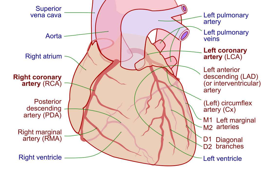
Low-molecular-weight heparin such as enoxaparin appears more in the pre-cath and acute coronary syndrome setting than as the intraprocedural agent, because it is harder to monitor with ACT and only partially reversible. Understanding how these agents protect coronary flow is easier once you can picture the vessels themselves.
Antiplatelets and DAPT: the stent's best friend
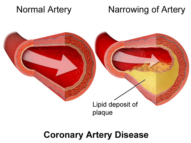
Anticoagulants handle the clotting cascade, but the coronary lesion itself is a platelet problem. When plaque ruptures — the event behind most heart attacks — platelets are the first responders, and a bare or drug-eluting stent is a platelet magnet until the vessel wall heals over it. That is why dual antiplatelet therapy (DAPT) is the cornerstone of PCI aftercare.
DAPT means two drugs working on two different pathways at once:
| Agent | Mechanism | Notes |
|---|---|---|
| Aspirin | Irreversibly blocks COX-1, cutting thromboxane A2 | Backbone of DAPT; a loading dose (often 162–325 mg chewed) is given before or during PCI |
| Clopidogrel | P2Y12 receptor blocker (prodrug) | Slower onset, variable metabolism; some patients are poor responders |
| Ticagrelor | P2Y12 blocker (reversible, not a prodrug) | Faster, more consistent; favored in many ACS pathways |
| Prasugrel | P2Y12 blocker (potent prodrug) | Strong platelet inhibition; avoided after prior stroke/TIA and in the elderly or low-weight |
The standard is aspirin plus one P2Y12 inhibitor, and the current guideline conversation centers on how long to continue the second agent. Historically twelve months after a drug-eluting stent was dogma; newer evidence supports shortening DAPT in high-bleeding-risk patients or, in some strategies, dropping aspirin and continuing potent P2Y12 monotherapy. This is genuinely evolving — the exact duration is individualized to ischemic versus bleeding risk, and the safest exam stance is that DAPT length is tailored, not fixed.
Glycoprotein IIb/IIIa inhibitors (abciximab historically, eptifibatide, tirofiban) block the final common pathway of platelet aggregation and are used intravenously as a bailout during high-thrombus-burden cases rather than routinely. They pack a strong antiplatelet punch and a matching bleeding risk. Because the coronary lesions being stented arise from the same disease process, it helps to understand coronary artery disease and how a myocardial infarction unfolds when a plaque ruptures. For the clinical distinction between the diagnostic study and the intervention, the piece on PCI versus cardiac catheterization is a useful companion.
Vasodilators: relaxing spasm and unmasking lesions
Coronary arteries are muscular tubes, and catheters irritate them. A wire tickling a vessel can trigger spasm — a transient narrowing that mimics a real blockage and can genuinely reduce flow. Intracoronary vasodilators relax that smooth muscle, restoring the vessel to its true diameter so the physician is treating anatomy, not artifact.
- Nitroglycerin is the reflexive first choice. Given directly into the coronary or intravenously, it dilates vessels (veins more than arteries at low dose), relieves spasm, and improves angiographic clarity. It also drops preload, so watch for hypotension. The dedicated article on nitroglycerin unpacks its mechanism and cautions in detail.
- Adenosine has a dual life in the lab: as a very short-acting AV-nodal blocker to break supraventricular tachycardia, and as the pharmacologic stress agent that induces maximal coronary hyperemia for fractional flow reserve (FFR) measurement. Its half-life is measured in seconds, which is exactly why it is safe to push.
- Calcium channel blockers such as verapamil or nicardipine, often mixed in the intracoronary "spasm cocktail," are potent at relieving refractory spasm and no-reflow.
- Sodium nitroprusside is an arterial and venous dilator reached for in hypertensive emergencies and no-reflow phenomena.
Vasodilators do more than improve pictures; they change the pressures the whole system runs on. Their effect on afterload connects directly to systemic vascular resistance, and the resulting shifts in flow are why understanding cardiac output matters when these drugs go in.
Vasoactive and emergency drugs: rescuing a crashing patient
The cath lab is where hearts occasionally stop, pressures collapse, and rhythms go haywire — often as a complication of the very disease being treated. The emergency drawer holds the agents that buy time. These are the vasoactive drugs, and the RCIS should know each one cold because in a code every second of hesitation counts.
| Drug | Primary action | Cath lab use |
|---|---|---|
| Epinephrine | Alpha + beta agonist | Cardiac arrest, profound hypotension, anaphylaxis |
| Norepinephrine | Strong alpha, some beta | First-line pressor for most shock states |
| Dopamine | Dose-dependent dopaminergic/beta/alpha | Hypotension with bradycardia |
| Dobutamine | Beta-1 inotrope | Low-output cardiogenic states to boost contractility |
| Atropine | Anticholinergic (vagolytic) | Symptomatic bradycardia, vagal reactions |
A classic scenario is the vagal reaction: during sheath insertion or removal a patient goes suddenly pale, bradycardic, and hypotensive. Atropine plus fluids is the textbook response. When the problem is pump failure rather than vagal tone, pressors and inotropes come out, and the physiology maps straight onto the four patterns described in the piece on shock hemodynamics. When drugs alone cannot hold the pressure, mechanical support such as the intra-aortic balloon pump is added.
Rhythm emergencies deserve their own reflexes. Ventricular arrhythmias during PCI call for antiarrhythmics like amiodarone or lidocaine and, when unstable, defibrillation — the electrical picture of which is covered in the guide to ventricular tachycardia. Because so many of these decisions are made off the monitor, fluency in reading the tracing is essential; brush up with the RCIS ECG guide and the walkthrough of STEMI ECG interpretation, since the acute coronary occlusion is often what brought the patient to the table.
Reversal agents: undoing the anticoagulation
Every drug that thins blood or blocks platelets can, at the wrong moment, become the enemy. Access-site hemorrhage, retroperitoneal bleeding, or perforation can turn a therapeutic anticoagulant into a life threat, and the reversal drawer is what turns the trade-off back toward safety.
- Protamine sulfate is the specific antidote for unfractionated heparin. It is a positively charged protein that binds the negatively charged heparin molecule and neutralizes it within minutes. It only partially reverses low-molecular-weight heparin and does nothing for bivalirudin. Protamine carries real risks of its own — hypotension, bradycardia, and anaphylactoid reactions, especially in patients previously exposed (including some diabetics on protamine-containing insulins) — so it is pushed slowly and thoughtfully.
- Direct thrombin inhibitors like bivalirudin have no reversal agent; management is supportive — stop the infusion, support the pressure, transfuse if needed, and wait for renal clearance. This is precisely why bivalirudin's short half-life is such an asset.
- Antiplatelet effects cannot be pharmacologically "reversed" in real time. Aspirin and the P2Y12 inhibitors act on the platelet for its lifespan, so significant bleeding may require platelet transfusion, and the effect wanes only as new platelets are made.
The broader point for the exam is directionality: know which agent each reversal drug targets, and know which agents have no antidote at all. That single distinction answers a surprising number of questions.
Contrast, sedation, and the supporting cast
Beyond the headline classes, several other agents round out a cath case, and the RCIS is expected to know their hazards.
Iodinated contrast media makes the coronaries visible under fluoroscopy, but it is not benign. It can cause allergic-type reactions and contrast-associated acute kidney injury, especially in patients with pre-existing renal disease, diabetes, or large dye loads. Hydration and dose limitation are the main defenses. The dedicated article on iodinated contrast media covers premedication and risk reduction.
Moderate (conscious) sedation — typically a benzodiazepine such as midazolam paired with an opioid such as fentanyl — keeps the patient comfortable and still without general anesthesia. The RCIS role here is heavy on monitoring: oxygen saturation, respiratory rate, and level of consciousness, because both drug classes depress respiration. Each has a reversal agent worth memorizing: flumazenil for benzodiazepines and naloxone for opioids.
Local anesthetic, usually lidocaine, numbs the access site at the groin or wrist before the sheath goes in. And a heparinized flush keeps the pressure-monitoring lines and catheters patent throughout. None of these are glamorous, but a missed sedation complication or an unrecognized contrast reaction can undo an otherwise flawless intervention.
Studying cath lab pharmacology for the RCIS
The registry does not ask you to be a pharmacist. It asks you to reason like the tech who anticipates the next syringe. A few habits make the material stick.
- Learn by job, not by name. When a drug appears, ask which of the five buckets it lives in. A stray drug name is far easier to place once you know whether it clots, un-clots, dilates, supports, or reverses.
- Pair each anticoagulant with its monitoring test and its antidote. Heparin → ACT → protamine. Bivalirudin → no specific antidote. That triad answers a huge share of questions.
- Anchor DAPT to the stent. Aspirin plus a P2Y12 inhibitor, duration individualized to bleeding versus ischemic risk. Know the four P2Y12 talking points and the one contraindication that trips people up (prasugrel after stroke/TIA).
- Rehearse the emergencies. Vagal reaction → atropine and fluids. Arrest → epinephrine. Refractory hypotension → norepinephrine. Bradycardia with hypotension → atropine, then pacing.
Round out your preparation with the interlocking calculators and rhythm skills the same patients demand: the hemodynamic calculator for pressure math, the cardiovascular anatomy guide for orientation, and targeted question sets in the RCIS hemodynamics practice and ECG practice banks. Repetition against real question stems is what converts these classes from a list into instinct.
Key takeaways
- Five jobs, not fifty drugs: cath lab medications either stop clotting, quiet platelets, dilate vessels, support the circulation, or reverse anticoagulation.
- Anticoagulants: heparin works indirectly through antithrombin, is monitored by ACT, and is reversed by protamine; bivalirudin is a direct thrombin inhibitor with no specific antidote — the go-to when HIT is a concern.
- DAPT is aspirin plus a P2Y12 inhibitor (clopidogrel, ticagrelor, or prasugrel), and its duration is individualized to bleeding versus ischemic risk under current, still-evolving guidance.
- Vasodilators like nitroglycerin and adenosine relieve spasm and unmask true lesion severity; adenosine also drives FFR and breaks SVT.
- Emergency vasoactive drugs — epinephrine, norepinephrine, dopamine, dobutamine, atropine — are the rescue kit for arrest, shock, and vagal reactions.
- Protamine reverses heparin; bivalirudin and antiplatelet agents have no fast reversal, so management is supportive.
- Don't forget the supporting cast: iodinated contrast, moderate sedation (reversed by flumazenil and naloxone), and local anesthetic each carry their own risks.
- This is educational content, not medical advice — real dosing and drug selection follow current guidelines and institutional protocol.
Practise cath-lab pharmacology
Test anticoagulants, antiplatelets, and reversal agents with explanations.
Practise Pharmacology →Frequently asked questions
What are the most common medications used in the cardiac cath lab?
The core classes are anticoagulants (heparin or bivalirudin), antiplatelets (aspirin plus a P2Y12 inhibitor such as ticagrelor, clopidogrel, or prasugrel), vasodilators (nitroglycerin, adenosine, calcium channel blockers), vasoactive emergency drugs (epinephrine, norepinephrine, atropine, dopamine, dobutamine), reversal agents (protamine), plus iodinated contrast and moderate sedation. Grouping them by the job they do makes them far easier to recall than a flat list.
What is DAPT and why is it used after a stent?
DAPT stands for dual antiplatelet therapy — aspirin combined with a P2Y12 inhibitor. After a coronary stent, the metal surface is a magnet for platelets until the vessel wall heals over it, so blocking two separate platelet pathways at once prevents stent thrombosis. Duration is individualized to a patient's bleeding versus ischemic risk under current, still-evolving guidelines rather than a single fixed number of months.
What is the difference between heparin and bivalirudin?
Heparin is an indirect anticoagulant that works by binding antithrombin, which then inactivates thrombin and factor Xa; it is monitored with the activated clotting time (ACT) and reversed with protamine. Bivalirudin is a direct thrombin inhibitor that binds thrombin itself, has a short predictable half-life, and has no specific antidote. Bivalirudin is often preferred in patients with high bleeding risk or a history of heparin-induced thrombocytopenia (HIT).
How is heparin reversed in the cath lab?
Unfractionated heparin is reversed with protamine sulfate, a positively charged protein that binds and neutralizes heparin within minutes. Protamine only partially reverses low-molecular-weight heparin and does not reverse bivalirudin. It is given slowly because it can cause hypotension, bradycardia, and anaphylactoid reactions, particularly in patients with prior protamine exposure.
Why is nitroglycerin given during a cardiac catheterization?
Nitroglycerin relaxes vascular smooth muscle, which relieves coronary spasm triggered by catheters and wires and dilates the vessel to its true diameter. This improves angiographic visualization and helps distinguish real stenosis from transient spasm. Because it also lowers preload, the main caution is hypotension, so blood pressure is watched closely when it is given.
What emergency drugs are kept ready in the cath lab?
The emergency kit typically includes epinephrine for cardiac arrest and profound hypotension, norepinephrine as a first-line pressor for shock, atropine for symptomatic bradycardia and vagal reactions, dopamine and dobutamine for low-output states, and antiarrhythmics such as amiodarone or lidocaine for ventricular arrhythmias. Reversal agents for sedation — flumazenil for benzodiazepines and naloxone for opioids — are also kept close.
What is a vagal reaction and how is it treated?
A vagal reaction is a sudden surge of parasympathetic tone, often during sheath insertion or removal, that causes pallor, bradycardia, hypotension, sweating, and sometimes nausea. The standard response is intravenous atropine to block the vagal effect on the heart plus fluids to restore blood pressure, with the patient often placed flat or head-down.
Can antiplatelet drugs be reversed quickly?
No — aspirin and P2Y12 inhibitors act on platelets for the platelet's lifespan, so there is no fast pharmacologic antidote. In serious bleeding, management is supportive and may include platelet transfusion, and the antiplatelet effect only fades as the body produces new platelets over several days.
Is contrast dye dangerous during a cath procedure?
Iodinated contrast is generally safe but carries two main risks: allergic-type reactions and contrast-associated acute kidney injury, especially in patients with pre-existing kidney disease, diabetes, or large dye volumes. Adequate hydration, limiting the contrast dose, and premedication in patients with prior reactions are the primary strategies to reduce those risks.
Sources & further reading
- Cardiovascular Credentialing International (CCI)
- American College of Cardiology
- American Heart Association
- MedlinePlus (U.S. National Library of Medicine)
External links are provided for reference; always confirm current details with the official source.