Radiation Safety in the Cardiac Cath Lab
Every case in the cardiac catheterization lab runs on ionizing radiation, and the technologist who understands how to control it protects the patient, the physician, and — over a whole career — themselves. This guide breaks down ALARA, the time-distance-shielding triad, the inverse square law, personal protective equipment, and dosimetry into the exam-ready, bench-ready essentials.
- Why radiation safety matters in the cath lab
- ALARA: the guiding principle
- Time, distance, and shielding: the protection triad
- The inverse square law explained
- Personal protective equipment (PPE)
- Dosimetry: monitoring your exposure
- Practical dose-reduction techniques
- Balancing patient and staff protection
- Key takeaways
Why radiation safety matters in the cath lab
Radiation safety in the cath lab is the disciplined, everyday practice of keeping X-ray exposure to patients and staff as low as reasonably achievable while still getting the images the procedure needs. Fluoroscopy is not a background detail of interventional cardiology — it is the imaging engine that makes the whole field possible. That also makes the cath lab one of the highest-occupational-dose environments in all of medicine.
The cardiovascular team stands beside the patient, case after case, year after year. Unlike a diagnostic radiographer who steps behind a wall, the invasive team works inside the room while the beam is on. Small doses that seem trivial on a single case accumulate. This is why regulatory bodies, professional societies, and every credentialing body treat radiation protection as a core competency rather than an optional extra. If you are new to the environment itself, our overview of what a cardiac cath lab is sets the scene for where all this equipment lives.
Radiation biology draws a useful line between two kinds of harm. Deterministic effects — such as skin erythema, epilation, or in extreme cases a radiation-induced skin injury — have a threshold dose and get worse as dose climbs; these are chiefly a patient concern during long, complex interventions. Stochastic effects — principally cancer induction — have no assumed threshold, and their probability rises with cumulative dose; these are the lifetime concern for staff. Good practice attacks both at once.
This article is educational and not medical advice. Dose thresholds, equipment specifications, and monitoring rules vary by device, jurisdiction, and institution; always follow current regulations and your local radiation-safety program.
ALARA: the guiding principle
ALARA stands for As Low As Reasonably Achievable, and it is the philosophical backbone of every radiation-protection decision in the lab. The word "reasonably" is doing real work in that phrase. ALARA does not demand zero dose — that would mean no imaging and no procedure. It demands that you take every practical, sensible step to minimize dose without compromising the clinical result.
ALARA is not a single rule but a mindset that plays out in dozens of small choices: keeping the fluoroscopy pedal down only when you are actually looking at the screen, using the lowest frame rate that still answers the clinical question, collimating tightly to the anatomy of interest, and storing the last fluoroscopy loop instead of shooting a fresh cine when a stored image will do. Each choice is minor on its own; together they can cut dose dramatically.
The three practical tools ALARA gives you to lower dose are the same three principles tested on every radiation-safety exam: time, distance, and shielding. The rest of this guide is essentially an expansion of how to use those three levers well, plus how to measure whether you are succeeding. Candidates working through our RCIS practice questions will see ALARA framed in scenario after scenario, because it is the concept every other radiation topic hangs from.
Time, distance, and shielding: the protection triad
The three primary principles of radiation protection are minimizing time, maximizing distance, and using shielding. They are listed together so often that they blur into a slogan, but each is a distinct, powerful lever, and understanding why each works turns memorization into intuition.
Time. Dose accumulates for exactly as long as the beam is on. Halve the fluoroscopy time and you halve the dose to everyone in the room, full stop. This is why beam-on time is displayed prominently and logged for every case, and why experienced operators pulse the pedal rather than holding it. Cine acquisition delivers far more dose per second than fluoroscopy, so minimizing unnecessary cine runs is part of the time discipline too.
Distance. Stepping back from the source is the most efficient protection available, because scatter radiation falls off steeply with distance — a relationship the inverse square law makes precise (see the next section). Even a single step away from the table meaningfully lowers a staff member's dose. The patient is the dominant source of scatter to the team, not the tube directly, so distance from the patient is what counts.
Shielding. Placing absorbing material between you and the scatter blocks a large fraction of it. This includes what you wear — lead aprons, thyroid collars, leaded glasses — and what hangs in the room: ceiling-suspended leaded acrylic shields, table-mounted skirts, and rolling barriers. Shielding is covered in depth in the PPE section below.
| Principle | How it lowers dose | Everyday application |
|---|---|---|
| Time | Dose is directly proportional to beam-on time | Pulse the fluoro pedal, use low frame rates, store fluoro loops, limit cine runs |
| Distance | Scatter falls off with the square of the distance | Step back when not at the table, use extension tubing, avoid leaning in unnecessarily |
| Shielding | Absorbing material attenuates scatter before it reaches you | Lead apron, thyroid collar, leaded glasses, ceiling and table shields |
The inverse square law explained
The inverse square law states that radiation intensity is inversely proportional to the square of the distance from the source — double your distance and the intensity drops to one quarter, not one half. This single relationship is why "distance" is such a potent protection tool, and it is a perennial exam favorite because it rewards understanding over rote recall.
Written out, the law is I₁/I₂ = (d₂)²/(d₁)², where I is intensity and d is distance. The practical takeaways are dramatic:
- Move from 1 meter to 2 meters and intensity falls to 25% (a factor of four).
- Move from 1 meter to 3 meters and intensity falls to about 11% (a factor of nine).
- Step in from 2 meters to 1 meter and you quadruple your exposure.
| Distance from source | Relative intensity | |
|---|---|---|
| 1 m (reference) | 100% | |
| 2 m | 25% | |
| 3 m | ~11% | |
| 4 m | ~6% |
Two nuances keep this honest at the bedside. First, the primary source of scatter to staff is the patient, so distances are measured from the patient, not from the X-ray tube in the abstract. Second, the inverse square law describes an idealized point source in air; real rooms have shielding, reflection, and beam geometry, so it is a guide to magnitude rather than a laboratory-exact prediction. Even so, its lesson is unambiguous: a small backward step buys a large reduction in dose, and it costs nothing.
Personal protective equipment (PPE)
Personal protective equipment in the cath lab means the lead-equivalent garments and barriers that absorb scatter before it reaches your body. No single piece is optional; together they form a layered defense, and each is aimed at the organs most vulnerable to radiation.
- Lead apron. The primary garment, typically 0.25 to 0.5 mm lead equivalent. Wrap-around styles protect the back for those who turn away from the source. A 0.5 mm apron attenuates the large majority of scatter at typical cath-lab energies. Aprons must be inspected regularly for cracks, because a break in the lead is a hidden hole in your protection.
- Thyroid collar. The thyroid is radiosensitive and sits unprotected above the apron line, so a wrap-around leaded collar is standard for anyone at the table.
- Leaded eyewear. The lens of the eye is the tissue most prone to radiation-induced cataract. Wraparound leaded glasses guard against this, and their importance grew when occupational eye-dose limits were tightened substantially in recent guidance.
- Ceiling-suspended and table-mounted shields. A leaded acrylic screen swung between the operator and the patient, plus a table skirt below, together block a striking amount of scatter to the head, neck, and lower body — often more than the apron alone.
| PPE item | Protects | Typical lead equivalent |
|---|---|---|
| Lead apron (wrap-around) | Torso, gonads, bone marrow | 0.25–0.5 mm Pb |
| Thyroid collar | Thyroid gland | 0.5 mm Pb |
| Leaded glasses | Lens of the eye (cataract risk) | ~0.75 mm Pb equivalent |
| Ceiling/table shields | Head, neck, lower body | Varies (leaded acrylic) |
Dosimetry: monitoring your exposure
Dosimetry is the measurement and recording of the radiation dose an individual worker actually receives, and it is what turns radiation safety from a hope into a verified fact. Every occupationally exposed staff member wears a dosimeter, and the readings are reviewed against regulatory limits.
The most common personal monitor is a badge worn on the outside of the lead apron at collar level. That collar badge estimates dose to unshielded areas — the head, neck, and eyes — which are the least protected. Many programs add a second badge worn under the apron at the waist to estimate protected trunk dose; comparing the two shows how well the apron is working. Pregnant workers who declare their pregnancy typically receive an additional fetal-dose badge worn at the waist under the apron, with a much lower monitoring threshold.
| Dosimeter placement | What it estimates |
|---|---|
| Collar badge (outside apron) | Dose to eyes, thyroid, and other unshielded tissue — the higher reading |
| Waist badge (under apron) | Protected deep-tissue/trunk dose |
| Fetal badge (declared pregnancy, under apron) | Estimated dose to the fetus, monitored to a stricter limit |
| Ring/extremity badge (select cases) | Dose to the hands during near-field work |
Dose is expressed in sieverts (Sv) for equivalent and effective dose, and because occupational doses are small, readings usually appear in millisieverts (mSv). Regulatory annual limits exist for whole-body effective dose, for the lens of the eye, and for extremities and skin; the eye-lens limit in particular was lowered dramatically in modern recommendations, which is the reason leaded eyewear moved from optional to expected. Exact numeric limits vary by jurisdiction, so always confirm the values your radiation-safety officer enforces rather than assuming a single global number.
Practical dose-reduction techniques
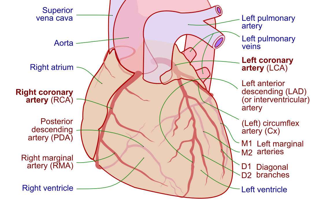
Beyond the big principles, a set of concrete, equipment-level habits separates a low-dose operator from a high-dose one. Most cost nothing and simply require attention. Because the coronary vessels are the frequent target of these procedures, being efficient with imaging directly limits exposure — a good moment to review the coronary artery anatomy you are trying to visualize so you spend less time hunting for it.
- Collimate tightly. Narrowing the beam to the anatomy of interest cuts scatter, lowers patient dose, and often improves image quality by reducing veiling glare.
- Use the lowest adequate frame rate. Dropping fluoroscopy from 15 to 7.5 frames per second can roughly halve dose with acceptable image quality for many tasks.
- Keep the image receptor close to the patient. A short air gap reduces the dose needed for a good image and lowers scatter.
- Minimize steep angulation and magnification. Steep (especially LAO cranial) projections and high magnification both raise dose and skin exposure.
- Vary the beam entry site on long cases. Rotating the gantry spreads the skin dose over a wider area, lowering the peak skin dose that drives deterministic injury.
- Store fluoroscopy loops instead of shooting new cine whenever a stored image answers the question.
These techniques protect the patient first and, because staff scatter comes from the patient, the team benefits simultaneously. That coupling is worth internalizing: nearly every action that lowers patient dose lowers your own. Candidates can test these ideas alongside the broader procedural picture in our comparison of PCI versus diagnostic cardiac catheterization, since intervention generally means longer beam-on time than a diagnostic study.
Balancing patient and staff protection
Radiation safety is often framed as protecting the worker, but the patient absorbs the primary beam and receives by far the largest dose in the room. The two goals are aligned more often than they conflict, yet a few distinctions are worth naming clearly.
For the patient, the dominant risk on long interventional cases is a deterministic skin injury from high cumulative skin dose. Modern systems track a reference air kerma or peak skin dose estimate, and many programs set a notification threshold that prompts the team to consider whether continuing is justified and to document follow-up for the patient's skin. Substudies and complex chronic-total-occlusion work are where this matters most. Recognizing when a case is trending toward a high skin dose is a shared team responsibility, not the operator's alone.
For staff, the concern is cumulative stochastic risk over a career plus lens-of-the-eye protection, addressed through the triad, PPE, and dosimetry already covered. A well-run lab folds both into one workflow: shorter beam-on time, tight collimation, and good shielding lower the patient's skin dose and the team's scatter at the same time.
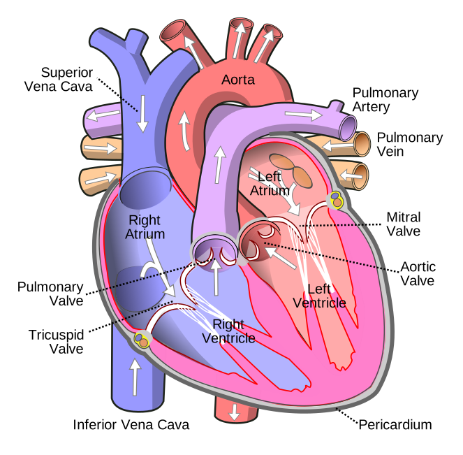
Context matters here too. Certain patients — those needing cardiac ablation, structural procedures, or repeat interventions for coronary artery disease — accrue radiation across multiple visits, so lifetime patient dose is part of the picture. Pediatric and pregnant patients warrant special caution and the most aggressive dose-minimization the clinical situation allows. When you connect radiation practice to the wider skill set — reading the ECG, understanding hemodynamics, and knowing the cardiac anatomy under the beam — you move faster and image less, which is itself a form of dose reduction.
Key takeaways
- ALARA — As Low As Reasonably Achievable — is the guiding principle: minimize dose without compromising the clinical result, chiefly by controlling fluoroscopy time.
- The three protection principles are time, distance, and shielding; nearly every dose-reduction question maps to one of them.
- The inverse square law means intensity drops with the square of distance — double your distance and dose falls to one quarter, which makes stepping back the highest-yield free habit.
- PPE is layered: a 0.25–0.5 mm lead apron, thyroid collar, leaded glasses for the radiosensitive eye lens, and ceiling/table shields that often block more scatter than the apron alone. Never fold an apron — hang it.
- Dosimetry verifies protection: a collar badge outside the apron estimates unshielded (eye/thyroid) dose, a waist badge under the apron estimates protected dose, and declared-pregnancy workers add a fetal badge. Dose is measured in sieverts (usually mSv).
- The patient absorbs the primary beam and faces deterministic skin-injury risk on long cases; staff face cumulative stochastic and eye-lens risk. Lowering patient dose usually lowers staff dose too.
- Practical habits — tight collimation, low frame rate, receptor close to patient, stored fluoro loops, varied beam angle — cut dose at little cost.
- This content is educational, not medical advice; exact dose limits and thresholds vary by jurisdiction and device, so follow your radiation-safety officer and current regulations.
Practise radiation safety
Test ALARA, dosimetry, and patient-care questions with instant feedback.
Practise Radiation Safety →Frequently asked questions
What does ALARA stand for in radiation safety?
ALARA stands for As Low As Reasonably Achievable. It is the core principle of radiation protection, meaning you take every practical and sensible step to keep radiation dose to patients and staff as low as possible without compromising the images the procedure requires. The word 'reasonably' is key — ALARA does not mean zero dose, it means no wasted dose. In the cath lab it plays out through short fluoroscopy times, low frame rates, tight collimation, and consistent shielding.
What are the three principles of radiation protection?
The three primary principles are time, distance, and shielding. Minimizing beam-on time reduces dose in direct proportion, since dose accumulates only while the beam is on. Maximizing distance from the source dramatically lowers exposure because scatter falls off with the square of the distance. Using shielding — lead aprons, thyroid collars, leaded glasses, and room shields — absorbs scatter before it reaches you. Almost every dose-reduction strategy comes down to one of these three levers.
How does the inverse square law apply to radiation safety?
The inverse square law states that radiation intensity is inversely proportional to the square of the distance from the source. If you double your distance from the source, the intensity drops to one quarter, not one half; triple the distance and it falls to about one ninth. This is why simply stepping back from the table is such an efficient and cost-free way to reduce occupational dose. Because the patient is the main source of scatter to staff, distance is measured from the patient.
What personal protective equipment is used in the cath lab?
Standard PPE includes a lead apron (usually 0.25 to 0.5 mm lead equivalent, ideally wrap-around), a thyroid collar to protect the radiosensitive thyroid gland, and leaded glasses to protect the lens of the eye from radiation-induced cataract. These are supplemented by ceiling-suspended leaded acrylic shields and table-mounted skirts, which together can block more scatter to the head, neck, and lower body than the apron alone. Lead aprons must be hung, never folded, to avoid cracking the lead.
Why should you never fold a lead apron?
Folding a lead apron repeatedly stresses the lead layer and creates cracks or gaps, and any break in the lead is a hidden hole in your protection where scatter passes straight through. Aprons should always be hung on proper racks. Programs also inspect aprons periodically with fluoroscopy or radiography to detect cracks, and any apron with significant damage is pulled from service because it can no longer be trusted to shield the wearer.
What is a dosimeter and where is it worn?
A dosimeter is a badge that measures and records the radiation dose an individual worker actually receives. The most common placement is a collar badge worn on the outside of the lead apron, which estimates dose to unshielded areas like the eyes and thyroid — the higher reading. Many programs add a second badge worn under the apron at the waist to estimate protected trunk dose, and declared-pregnant workers wear an additional fetal-dose badge under the apron. Radiation dose is measured in sieverts, usually reported in millisieverts.
What is the difference between deterministic and stochastic radiation effects?
Deterministic effects have a threshold dose and become more severe as dose increases; examples include skin reddening, hair loss, and cataracts, and they are mainly a patient concern during long, high-dose interventional cases. Stochastic effects, chiefly cancer induction, have no assumed threshold — their probability rises with cumulative dose, though severity does not — and they are the main lifetime concern for staff. Good radiation practice targets both by minimizing every avoidable exposure.
How can fluoroscopy dose be reduced during a procedure?
The biggest lever is reducing beam-on time by keeping your foot off the pedal unless you are actually looking at the screen. Other proven techniques include collimating tightly to the anatomy of interest, using the lowest adequate frame rate (for example, dropping from 15 to 7.5 frames per second), keeping the image receptor close to the patient, minimizing steep angulation and magnification, varying the beam entry angle on long cases to spread skin dose, and storing fluoroscopy loops instead of shooting new cine. These lower patient dose and staff scatter at the same time.
Who receives more radiation dose, the patient or the staff?
The patient absorbs the primary X-ray beam and receives by far the largest dose in the room, with the main risk on long interventional cases being a deterministic skin injury from high cumulative skin dose. Staff receive only scattered radiation, which is a small fraction of the patient's dose, but they accumulate it across many cases over a career, so their concern is long-term stochastic risk and protection of the eye lens. Importantly, most actions that lower the patient's dose also lower the scatter reaching the team.
Sources & further reading
- Cardiovascular Credentialing International (CCI)
- American College of Cardiology
- American Heart Association
- MedlinePlus (U.S. National Library of Medicine)
External links are provided for reference; always confirm current details with the official source.