PCI vs Cardiac Catheterization
People often use "PCI" and "cardiac catheterization" as if they mean the same thing, but they describe two different jobs done in the same room — one looks, the other fixes. Understanding where the diagnostic study ends and the interventional treatment begins is one of the most useful mental models in all of cardiovascular care.
- Same lab, different jobs
- Diagnostic vs interventional: the core distinction
- What happens during a diagnostic catheterization
- What happens during PCI: angioplasty and stenting
- When each procedure is done
- Can both happen in one visit?
- How the team decides a blockage needs treating
- Risks, recovery, and the RCIS team
- Key takeaways
Same lab, different jobs
Walk into a modern cardiac cath lab and you would struggle to tell a diagnostic case from an interventional one just by looking. The X-ray equipment, the sterile drapes, the monitors, and the catheters lined up on the table are largely the same. What differs is the intent — and that difference is the whole point of this article.
Cardiac catheterization is the broad, umbrella term. It means threading a thin, hollow tube (a catheter) through a blood vessel into the heart or its arteries to gather information. When that information-gathering is the entire goal, the study is diagnostic. Percutaneous coronary intervention (PCI) is a specific therapeutic subset — using catheters not to measure or picture the problem, but to physically treat a blocked coronary artery with a balloon and, almost always, a stent.
Put simply: every PCI begins with a catheterization, but not every catheterization becomes a PCI. The catheter is the shared tool; the question is whether you are collecting data or delivering treatment. This piece is written for RCIS candidates, cardiovascular technologists, and curious patients. It is educational and not a substitute for medical advice — decisions about your own care belong with your cardiologist.
Diagnostic vs interventional: the core distinction
The cleanest way to hold this in your head is the difference between a diagnostic procedure and an interventional one. A diagnostic catheterization answers questions. An interventional procedure — PCI being the headline example — solves the problem it finds.
During a diagnostic study, the operator injects contrast dye and records X-ray movies (an angiogram) to see whether the coronary arteries are narrowed, and by how much. They may also measure pressures inside the heart chambers, sample oxygen levels, and calculate flow. Nothing about the artery is changed. The patient leaves with answers — a map of their disease — and a plan.
An interventional procedure takes that map and acts on it. If a diagnostic angiogram reveals a tight, flow-limiting blockage, the team can, often in the same sitting, advance a guidewire across the lesion, inflate a balloon to crack it open (angioplasty), and deploy a stent to hold the vessel open. The narrowing that was merely observed a moment ago is now treated.
| Feature | Diagnostic catheterization | PCI (interventional) |
|---|---|---|
| Primary goal | Find and measure disease | Treat a specific blockage |
| What is done | Inject dye, image, measure pressures | Balloon angioplasty + stent placement |
| Changes the artery? | No | Yes |
| Typical duration | 20–45 minutes | 45–90+ minutes |
| Blood thinners | Minimal | Heparin/antiplatelets essential |
| Recovery | A few hours | Hours to overnight |
What happens during a diagnostic catheterization
A diagnostic cardiac catheterization is a remarkably choreographed study. After a local anesthetic numbs the access site — most often the radial artery at the wrist, sometimes the femoral artery in the groin — the operator inserts a short tube called a sheath. Through it, catheters are advanced under live X-ray guidance up to the heart.
From there, the study can go in several directions depending on the clinical question:
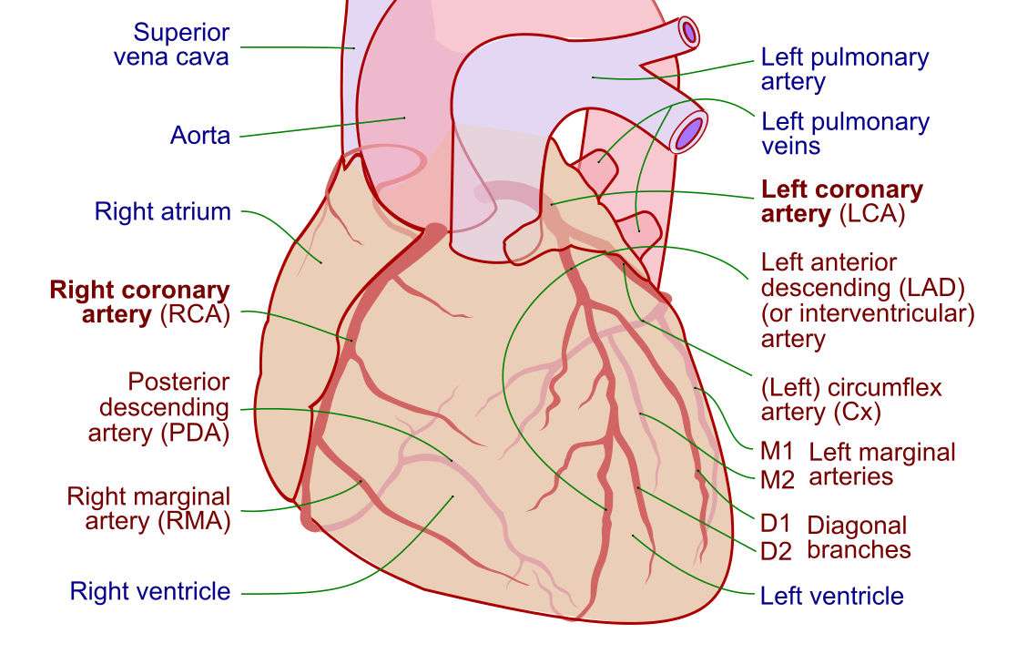
- Coronary angiography — the most common component. Contrast dye is injected into the left and right coronary arteries and filmed, producing the classic branching "tree" images that reveal narrowings.
- Left heart catheterization — a catheter crosses into the left ventricle to measure its pressures and, sometimes, perform a ventriculogram that shows how well the chamber squeezes and estimates the ejection fraction.
- Right heart catheterization — using a balloon-tipped Swan-Ganz catheter to record right atrial, right ventricular, pulmonary artery, and wedge pressures, and to calculate cardiac output.
These pressure measurements are where a firm grasp of hemodynamic principles pays off. The waveforms and numbers collected here feed calculations like cardiac index, stroke volume, and systemic vascular resistance — the vocabulary of the RCIS exam and the daily work of the lab. Throughout, the patient is awake but sedated, monitored by continuous ECG and pressure tracings.
Crucially, at the end of a purely diagnostic case, the arteries are exactly as they were at the start. The value delivered is knowledge: a precise, individualized picture of the patient's coronary anatomy and heart function that guides every decision to follow.
What happens during PCI: angioplasty and stenting
PCI picks up where diagnosis leaves off. Once a significant blockage is confirmed — often with the help of physiologic testing, discussed below — the operator commits to treating it. The steps build logically on the diagnostic access already in place.
- Anticoagulation. Because catheters and wires will now spend extended time inside a coronary artery, the patient is given heparin and potent antiplatelet drugs to prevent clot formation. This is a defining difference from diagnostic work.
- Crossing the lesion. A hair-thin guidewire is steered across the narrowing — the trickiest, most skill-dependent moment of the case.
- Balloon angioplasty. A deflated balloon rides over the wire to the blockage and is inflated, compressing plaque and stretching the vessel open.
- Stent deployment. A tiny metal-mesh scaffold — nearly always a drug-eluting stent that slowly releases medication to prevent re-narrowing — is expanded against the artery wall, where it stays permanently.
- Confirmation. Fresh contrast injections confirm restored flow. Imaging or pressure wires may verify the result.
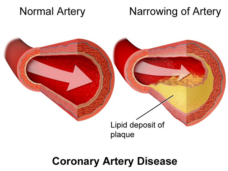
The term angioplasty technically refers just to the balloon step; the modern combination of balloon plus stent is what most clinicians mean when they say PCI. Balloon-only angioplasty is now reserved for special situations, because bare balloon dilation alone carries a higher chance of the vessel snapping shut or re-narrowing. Understanding the coronary artery anatomy being treated — which vessel, how many branches, how much muscle it feeds — shapes every one of these choices.
When each procedure is done
Knowing when each is chosen is where students often stumble, so it is worth being concrete. The two procedures answer different clinical needs, and sometimes both happen back-to-back.
A diagnostic catheterization is typically done when clinicians need to define coronary anatomy or heart function, such as:
- Persistent chest pain or an abnormal stress test suggesting coronary artery disease that non-invasive tests could not fully clarify.
- Evaluating unexplained heart failure or a low ejection fraction to see whether blocked arteries are the cause.
- Assessing valve disease, congenital defects, or pressures in conditions like pulmonary hypertension.
- Working up a patient before heart surgery to map the coronary arteries.
PCI is done when a blockage is both significant and treatable by catheter, most importantly:
- ST-elevation myocardial infarction (STEMI) — the clearest indication. Guidelines call for emergency primary PCI to reopen the culprit artery as fast as possible; the door-to-balloon clock is literally muscle-saving. A confident read of the STEMI ECG is what triggers this pathway.
- Non-ST-elevation acute coronary syndromes — often treated with PCI within hours to a day, depending on risk.
- Stable angina that keeps limiting a patient despite good medical therapy, when a suitable lesion is present.
An important, evolving nuance: for stable coronary disease, large trials have tempered the old "see a blockage, stent a blockage" reflex. Optimal medications often perform as well as stenting for preventing hard outcomes in stable patients, so PCI in that setting is increasingly reserved for relieving symptoms that medicine cannot. The evidence here continues to be refined, and decisions are individualized.
Can both happen in one visit?
Yes — and this "ad hoc" PCI is one of the most elegant features of modern cardiology. Because the diagnostic study and the intervention share the same access, X-ray system, and skill set, a cardiologist who finds a treatable, high-grade blockage during a diagnostic angiogram can often move straight into PCI without a second procedure on another day.
This is enormously efficient. It spares the patient a repeat hospital visit, a second arterial puncture, and a second dose of contrast and radiation. In a STEMI, it is not merely convenient — it is the standard of care, because every minute of delay costs heart muscle.
But it is not automatic. Several factors prompt a deliberate pause between diagnosis and treatment:
- Complex or multi-vessel disease, where the best strategy might be bypass surgery rather than stenting, warrants a "heart team" discussion first.
- Left main disease or intricate anatomy may favor a staged, carefully planned approach.
- Patient stability and consent — a stable patient deserves the chance to weigh options rather than being committed to a stent under sedation.
So while the tools make same-visit treatment possible, good practice means treating on the spot when the case is clear and urgent, and stepping back to plan when it is complex. The catheter's versatility is a gift, but judgment decides when to use it.
How the team decides a blockage needs treating
Not every narrowing seen on an angiogram deserves a stent. A blockage that looks alarming on film may not actually be starving the muscle downstream, and treating it needlessly exposes the patient to risk without benefit. This is where physiology enters — and it is a favorite topic on cardiovascular exams.
To settle borderline cases, interventionalists thread a pressure-sensing guidewire across the lesion and measure fractional flow reserve (FFR) or similar resting indices. By comparing pressure before and after the narrowing (usually while dilating the microvasculature with a drug), FFR quantifies whether the lesion truly limits flow. A value below the accepted cutoff means the blockage is functionally significant and likely benefits from PCI; a value above it means medical therapy is often the wiser path.
This physiologic gatekeeping reflects a broader shift: from treating what a stenosis looks like to treating what it does. The same hemodynamic reasoning that underlies FFR runs through the hemodynamic calculations used elsewhere in the lab and through classic measurements like the Fick cardiac output. Intravascular imaging — ultrasound or optical coherence tomography — can add anatomic detail, sizing the vessel and checking that a stent is fully expanded.
Risks, recovery, and the RCIS team
Both procedures are generally safe in experienced hands, but their risk profiles differ in proportion to how much they do. A diagnostic catheterization is lower-risk: the main concerns are bleeding or bruising at the access site, contrast reactions, kidney stress from the dye, and — rarely — vessel injury or arrhythmia. Most patients go home the same day after a few hours of monitoring.
PCI carries everything diagnostic work does, plus risks tied to manipulating a diseased artery: the treated vessel can abruptly close, small clots or debris can travel downstream, and, uncommonly, the artery can be perforated. Because a permanent stent is placed, patients must take dual antiplatelet therapy — typically aspirin plus a second agent — for a defined period to keep the stent from clotting. Skipping these drugs early is genuinely dangerous. Detailed patient education, often drawing on the lab's cath lab medications knowledge, is part of a safe discharge.
None of this happens solo. The cath lab is a team sport, and the cardiovascular technologist and RCIS-credentialed staff are central to it — scrubbing in, managing hemodynamic monitoring, operating imaging and physiology equipment, handling devices, and watching the ECG and pressures for the first sign of trouble. Solid command of both cardiac anatomy and real-time waveform interpretation is exactly what the credential certifies, and exactly what keeps these procedures safe.
Key takeaways
- Cardiac catheterization is the umbrella term for threading catheters into the heart; when the goal is only to gather information, it is diagnostic.
- PCI (percutaneous coronary intervention) is the interventional subset — balloon angioplasty plus a stent to physically treat a blockage.
- The core distinction: diagnostic catheterization looks and measures and changes nothing; PCI treats and permanently alters the artery.
- Angioplasty + stent is what most people mean by PCI; the balloon opens the vessel and the drug-eluting stent holds it open.
- Diagnostic studies are chosen to define anatomy and function; PCI is chosen for significant, treatable blockages — above all, emergency primary PCI for STEMI, where time is muscle.
- Both can happen in one visit ("ad hoc" PCI), but complex or multi-vessel disease may warrant a heart-team pause first.
- Physiology such as FFR helps decide whether a narrowing truly needs treating — function matters more than appearance.
- PCI carries added risks and requires dual antiplatelet therapy; the RCIS-credentialed team is essential to performing both procedures safely.
- This article is educational and not medical advice; discuss your own care with your cardiologist.
Frequently asked questions
Is PCI the same as cardiac catheterization?
No. Cardiac catheterization is the broad term for inserting a catheter into the heart or its arteries, usually to diagnose problems. PCI (percutaneous coronary intervention) is a specific treatment done through a catheter — using a balloon and stent to open a blocked coronary artery. Every PCI involves catheterization, but most diagnostic catheterizations are not PCIs.
What is the difference between diagnostic and interventional cardiac catheterization?
A diagnostic catheterization gathers information: it uses contrast dye and X-rays to picture the arteries and measures pressures inside the heart, without changing anything. An interventional procedure, such as PCI, treats what it finds — for example, opening a narrowed artery with a balloon and stent. Diagnostic is the camera; interventional is the repair.
Is angioplasty the same as PCI?
Angioplasty technically refers to inflating a balloon to open a narrowed artery. PCI is the broader modern term that usually includes angioplasty plus placing a stent. In everyday practice, when people say PCI they almost always mean balloon angioplasty followed by a drug-eluting stent, since stenting greatly reduces the chance the artery re-narrows.
Can diagnostic catheterization and PCI be done at the same time?
Yes. Because both use the same catheter access and X-ray equipment, a cardiologist who finds a significant, treatable blockage during a diagnostic angiogram can often proceed directly to PCI in the same session — called ad hoc PCI. In a STEMI heart attack this is standard. For complex or multi-vessel disease, the team may pause to plan or consider bypass surgery first.
When is PCI needed instead of just a diagnostic cath?
PCI is needed when a coronary artery is significantly blocked and the blockage can be treated with a catheter. The clearest case is a STEMI heart attack, which calls for emergency primary PCI. It is also used for other acute coronary syndromes and for stable angina that keeps limiting a patient despite medication. A diagnostic cath alone is enough when the goal is simply to map disease or assess heart function.
Does a diagnostic catheterization always lead to a stent?
No. Many diagnostic catheterizations show no significant blockages, or narrowings that are better managed with medication and lifestyle changes rather than a stent. Physiologic testing such as FFR helps decide whether a given blockage actually limits blood flow enough to warrant PCI. Appearance on the angiogram alone does not determine treatment.
How long does PCI take compared with a diagnostic cath?
A diagnostic cardiac catheterization typically takes about 20 to 45 minutes, and most patients go home the same day after a few hours of monitoring. PCI usually takes longer — often 45 to 90 minutes or more depending on complexity — and may involve a longer observation period, sometimes overnight, because a stent has been placed and blood thinners are used.
What is FFR and why does it matter in PCI?
FFR (fractional flow reserve) is a measurement taken with a pressure-sensing wire to see whether a narrowing truly limits blood flow to the heart muscle. A value below the accepted cutoff means the blockage is functionally significant and likely benefits from PCI, while a higher value suggests medication is the better choice. FFR helps avoid stenting blockages that only look severe but do not restrict flow.
Do you need blood thinners after PCI?
Yes. After PCI with a stent, patients take dual antiplatelet therapy — usually aspirin plus a second antiplatelet drug — for a defined period to stop a clot from forming on the new stent. Stopping these medications early can be dangerous and cause stent thrombosis. Diagnostic catheterization alone generally does not require this ongoing therapy.
Sources & further reading
- Cardiovascular Credentialing International (CCI)
- American College of Cardiology
- American Heart Association
- MedlinePlus (U.S. National Library of Medicine)
External links are provided for reference; always confirm current details with the official source.