Coronary Angiography vs Cardiac Catheterization
"Coronary angiography" and "cardiac catheterization" get used as if they mean the same thing — and in casual conversation they nearly do — but they are not identical. One is the broad family of procedures; the other is a specific test that lives inside it.
- Coronary angiography vs cardiac catheterization: the quick answer
- Are coronary angiography and cardiac catheterization the same thing?
- What cardiac catheterization actually includes
- What coronary angiography actually shows
- Key differences at a glance
- How the combined procedure works, step by step
- When each procedure is used clinically
- Risks, contrast, and safety considerations
- RCIS exam focus: how this shows up on the test
- Key takeaways
Coronary angiography vs cardiac catheterization: the quick answer
Here is the short version, and it is the answer most people are actually looking for: cardiac catheterization is the umbrella term for any procedure in which a thin, flexible tube — a catheter — is threaded through a blood vessel into the heart. Coronary angiography is one specific thing you can do once that catheter is in place: inject contrast dye into the coronary arteries and film them with X-ray to look for blockages.
So the relationship is not "A versus B" so much as "B is a part of A." Every coronary angiogram is a cardiac catheterization. But not every cardiac catheterization includes coronary angiography — you might catheterize the right side of the heart to measure pressures and never touch the coronary arteries at all.
The confusion is understandable. In everyday practice, when a patient is sent for a "cath," coronary angiography is usually the main event, so the words blur together. But on the RCIS exam, and in a well-run cath lab, the distinction matters. It tells you which chambers and vessels are being studied, what equipment is on the table, and what the report is meant to answer.
One-line memory hook: Catheterization is the journey (getting the catheter to the heart). Angiography is one destination (dyeing and filming the coronary arteries).
Are coronary angiography and cardiac catheterization the same thing?
No — but they overlap so heavily that treating them as synonyms rarely causes harm at the bedside. The precise way to say it: coronary angiography is a subset of cardiac catheterization. It is what you get when the catheter is used to opacify and image the coronary circulation.
Think of cardiac catheterization as a toolkit and the catheter as the tool that reaches the heart. Once you are there, you can do several different jobs:
- Coronary angiography — inject dye into the left and right coronary arteries to map atherosclerotic disease.
- Left heart catheterization — cross the aortic valve into the left ventricle to measure pressures and, often, perform a ventriculogram.
- Right heart catheterization — advance a balloon-tipped catheter through the right atrium, right ventricle, and pulmonary artery to measure filling pressures, cardiac output, and vascular resistance.
- Interventions — angioplasty, stenting, valve procedures, and biopsy all begin as catheterizations.
Coronary angiography is simply the most commonly performed of these jobs, which is why it dominates the conversation. If you want the deeper mechanics of what the catheter does once inside the heart, our overview of what happens inside a cardiac cath lab walks through the workflow end to end.
What cardiac catheterization actually includes
Because it is the broader category, cardiac catheterization can answer a wide range of clinical questions — many of which have nothing to do with the coronary arteries. That breadth is the whole point of keeping it as a distinct, larger term.
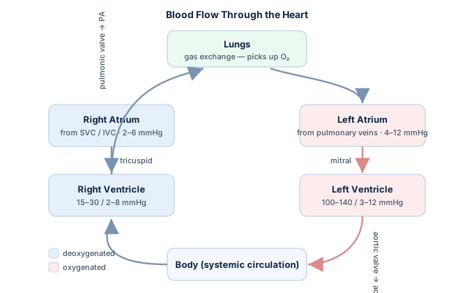
A full diagnostic catheterization can involve any combination of the following, depending on what the cardiologist needs to know:
| Component | What it measures or shows | Coronary arteries involved? |
|---|---|---|
| Coronary angiography | Location and severity of coronary blockages | Yes — this is the point |
| Left ventriculography | Ejection fraction, wall motion, mitral regurgitation | No |
| Right heart cath | RA, RV, PA, and wedge pressures; cardiac output | No |
| Aortic "pullback" | Pressure gradient across the aortic valve | No |
| Shunt run | Oxygen saturations to detect intracardiac shunts | No |
Notice that most of what catheterization can do lives in the pressure-and-flow world, not the coronary-anatomy world. The hemodynamic side leans on concepts like cardiac output, stroke volume, and systemic vascular resistance — the numbers a right heart study is designed to produce. If waveform interpretation is your weak spot, the RCIS hemodynamics guide is the fastest way to shore it up.
Interventional catheterizations extend the toolkit further, layering therapy on top of diagnosis. The line between diagnostic angiography and treatment is spelled out in our comparison of PCI versus cardiac catheterization, which is worth reading alongside this one.
What coronary angiography actually shows
Coronary angiography has a narrow, well-defined job: to reveal the anatomy of the coronary arteries in high enough detail to guide treatment decisions. Contrast dye is injected directly into the coronary ostia, and rapid X-ray imaging (cineangiography) records the dye filling — or failing to fill — each vessel.
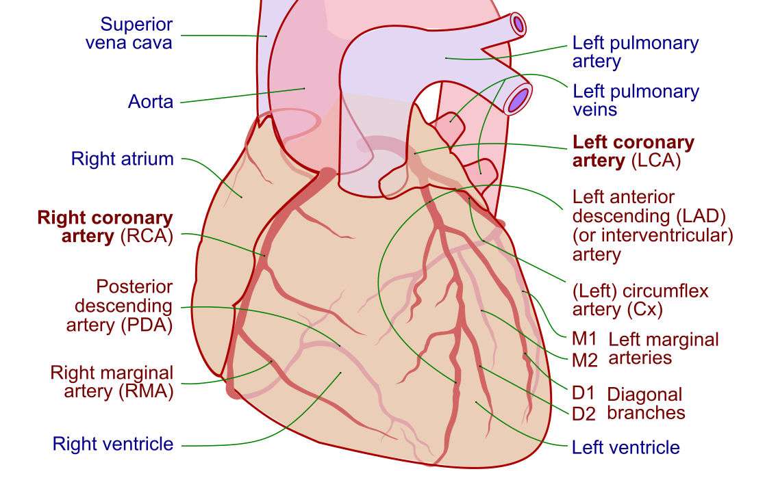
Specifically, a coronary angiogram answers questions like:
- Where are the blockages? Which artery — left main, left anterior descending, circumflex, or right coronary — and which segment.
- How severe are they? Stenoses are graded by percent diameter narrowing; a lesion above roughly 70 percent (or 50 percent in the left main) is generally considered significant.
- Is a lesion functionally important? Angiographic severity does not always equal physiologic significance, so borderline lesions are increasingly assessed with a pressure wire (fractional flow reserve, FFR) rather than eyeballed alone — a shift reflected in current revascularization guidance.
- What is the treatment path? The map determines whether a patient goes to stenting, bypass surgery, or medical therapy.
Firm knowledge of the vessels themselves makes every angiogram easier to read. Our detailed page on coronary artery anatomy and the broader cardiovascular anatomy guide cover the branching patterns and dominance you will be expected to recognize on film. When angiography reveals disease, the clinical context usually traces back to coronary artery disease or an evolving myocardial infarction.
Important caveat: Angiography shows the lumen — the open channel dye can flow through. It underestimates plaque burden in the wall itself, because arteries can remodel outward to preserve the lumen even as disease builds. That is why intravascular imaging (IVUS, OCT) is sometimes added.
Key differences at a glance
When you strip away the overlap, the core differences come down to scope, purpose, and what the final report contains. This table is the one to memorize.
| Feature | Cardiac catheterization | Coronary angiography |
|---|---|---|
| Scope | Broad umbrella procedure | One specific test within it |
| Primary target | Any chamber, valve, or vessel of the heart | The coronary arteries only |
| Main output | Pressures, flows, saturations, and/or images | X-ray images of coronary anatomy |
| Uses contrast dye? | Sometimes (not for a pure right heart pressure study) | Always — dye is the whole method |
| Answers | How is the heart pumping and filling? | Where and how bad are the blockages? |
| Relationship | Contains angiography | Is contained by catheterization |
The single cleanest distinction: catheterization is defined by the method (a catheter reaches the heart), while angiography is defined by the imaging technique (contrast plus X-ray). You can catheterize without imaging vessels, but you cannot perform coronary angiography without first catheterizing.
How the combined procedure works, step by step
In the real world these two rarely happen in isolation. A typical "diagnostic cath" bundles catheterization and coronary angiography — and sometimes ventriculography — into a single session lasting well under an hour. Understanding the flow helps cement why the terms overlap.
- Access. The operator numbs and punctures an artery — most often the radial artery at the wrist, increasingly preferred over the femoral approach because of lower bleeding risk in current practice. A sheath holds the door open.
- Navigation. Under X-ray (fluoroscopy), a diagnostic catheter is advanced up the aorta to the heart. No dye is needed yet — this is pure catheterization.
- Engagement. The catheter tip is seated in the opening of a coronary artery.
- Angiography. Contrast is hand-injected while the X-ray camera films from multiple angles. This step — and only this step — is the coronary angiogram.
- Optional add-ons. A pigtail catheter may cross into the left ventricle for a ventriculogram (giving an ejection fraction), or a pressure wire may interrogate a borderline lesion.
- Closure. The sheath comes out; the site is sealed with pressure or a closure device.
Throughout, the patient stays awake under light sedation, and the team manages anticoagulation and vasoactive drugs from the cath lab medication shelf. If a blockage found on angiography needs treatment, the case can convert to an intervention in the same sitting — which is where a device like the intra-aortic balloon pump or a Swan-Ganz catheter may enter the picture for a sicker patient.
This article is educational and written for exam preparation and general understanding — it is not medical advice. Procedural decisions belong to the treating cardiology team.
When each procedure is used clinically
Because their scopes differ, the clinical triggers differ too. Coronary angiography is ordered when the question is specifically about the coronary arteries; broader catheterization is ordered when the question is about pressures, flows, valves, or shunts.
Coronary angiography is typically indicated for:
- Suspected or known coronary artery disease with limiting symptoms.
- Acute coronary syndromes — an unstable angina or heart attack presentation, where a STEMI on the ECG often sends a patient straight to the lab.
- Abnormal stress testing or evolving symptoms despite medical therapy.
- Pre-operative assessment before valve or other cardiac surgery.
Broader (often right heart) catheterization is used for:
- Working up unexplained heart failure or pulmonary hypertension.
- Assessing shock states and guiding therapy — the pressure patterns of shock hemodynamics and cardiac tamponade are classic exam material.
- Quantifying valve gradients, such as the aortic valve area in suspected stenosis.
- Calculating derived numbers like cardiac index at the bedside.
A quick surface ECG is part of nearly every cath, and knowing your rhythms cold — from a benign one on the ECG interpretation primer to a dangerous run of ventricular tachycardia — keeps you oriented when the monitor changes mid-case.
Risks, contrast, and safety considerations
Both procedures share a strong safety record, but their risk profiles are not identical — and the difference traces directly back to that dye. Coronary angiography always uses iodinated contrast, so it carries risks a pure pressure-measuring catheterization does not.
- Contrast-related risk. Iodinated dye can provoke allergic-type reactions and can stress the kidneys (contrast-associated acute kidney injury), a concern that scales with pre-existing kidney disease and dye volume. A right heart pressure study using no dye sidesteps this entirely.
- Radiation. Angiography and interventions use fluoroscopy; longer, more complex cases mean more dose. Modern labs minimize this, but it is real.
- Access-site and vascular risk. Shared by all catheterizations — bleeding, hematoma, and rarely vessel injury. Radial access has lowered this meaningfully.
- Rare major events. Stroke, heart attack, arrhythmia, and dissection are uncommon but possible in any catheterization.
Exam tip: If a question stem emphasizes kidney function, dye allergy, or metformin, it is steering you toward angiography-specific (contrast) concerns — not the generic catheterization risks that apply even without dye.
The overall message from current guidance is reassuring: for a stable patient having an elective study, the risk of a serious complication sits well below one percent. Evidence on optimal contrast agents, closure strategies, and radial-first protocols continues to evolve, so specifics can shift as new data lands. Vasoactive and antiplatelet agents used to manage these risks are covered in our review of medications used in the cath lab, and the role of the imaging team is detailed on the cardiovascular ultrasound technologist page.
RCIS exam focus: how this shows up on the test
Registry writers love this distinction precisely because casual usage blurs it. Expect at least one item that hinges on knowing that angiography is a component, not a synonym. A few patterns to watch for:
- Definitional swaps. A stem may describe a right heart study measuring wedge pressure and ask what it is — the wrong-but-tempting answer is "coronary angiography." It is catheterization without angiography.
- Contrast logic. Any question tying dye, kidneys, or allergies to the procedure is testing whether you know angiography always uses contrast while some catheterizations do not.
- Hemodynamic integration. You will often need to pair the anatomy concept with numbers — pull the pressure and flow relationships from the hemodynamics guide and practice them against the hemodynamics question bank.
- Rhythm awareness. ECG never fully leaves the picture; the rhythm interpretation refresher and the ECG strip drills keep that muscle warm.
Work these alongside the ECG guide and the free hemodynamic calculator so the vocabulary and the math reinforce each other. The goal is to reach the exam able to say, in one breath, exactly how these two terms relate.
Key takeaways
- Cardiac catheterization is the umbrella; coronary angiography is one specific test performed within it.
- Every coronary angiogram is a catheterization, but many catheterizations (especially right heart studies) include no angiography at all.
- Angiography always uses contrast dye and X-ray to map coronary blockages; a pure pressure study may use neither.
- Catheterization is defined by method (a catheter reaches the heart); angiography is defined by imaging technique (dye plus fluoroscopy).
- Clinical trigger differs: angiography answers "where are the blockages," while broader catheterization answers "how is the heart filling and pumping."
- Angiography-specific risks — contrast reactions and kidney stress — are the exam's favorite way to separate the two.
- For the RCIS, know the definitions cold and pair them with hemodynamic numbers and basic rhythm recognition.
- This content is educational and not a substitute for professional medical advice.
See where it all happens
Learn what a cardiac cath lab is and who is in the room.
What Is a Cardiac Cath Lab →Frequently asked questions
Are coronary angiography and cardiac catheterization the same thing?
No, though they overlap heavily. Cardiac catheterization is the broad procedure of threading a catheter into the heart; coronary angiography is one specific test — injecting dye into the coronary arteries and filming them — performed during a catheterization. Every coronary angiogram is a catheterization, but not every catheterization includes angiography.
What is the main difference between the two?
Scope. Catheterization is the umbrella method defined by getting a catheter to the heart, and it can measure pressures, flows, and saturations anywhere in the heart. Coronary angiography is narrowly focused on imaging the coronary arteries with contrast dye and X-ray.
Does coronary angiography always use contrast dye?
Yes. Contrast is the entire method — the dye is what makes the coronary arteries visible on X-ray. By contrast, a right heart catheterization that only measures pressures may use no dye at all, which is a key clinical and exam distinction.
What does coronary angiography show that catheterization alone does not?
It reveals the anatomy of the coronary arteries: the location, severity, and pattern of blockages. A catheterization done purely for hemodynamics gives you pressures and flows but no picture of the coronary vessels themselves.
Can you have cardiac catheterization without coronary angiography?
Yes. A right heart catheterization to work up heart failure or pulmonary hypertension threads a catheter through the right atrium, right ventricle, and pulmonary artery to measure pressures and cardiac output — without ever imaging the coronary arteries.
Which procedure is riskier?
They share a strong safety record, but coronary angiography carries the added risks of contrast dye (allergic reactions and kidney stress) and fluoroscopic radiation. A pure pressure-measuring catheterization avoids the dye-related risks. For stable elective patients, serious complications occur in well under one percent of cases.
Why do people use the terms interchangeably?
Because in everyday practice, when a patient is sent for a "cath," coronary angiography is usually the main event. The words blur together in casual speech, but the technical distinction still matters for reporting, equipment, and exams.
Is coronary angiography the same as PCI or angioplasty?
No. Angiography is diagnostic — it only images the arteries. PCI (percutaneous coronary intervention), including angioplasty and stenting, is the therapeutic step that treats a blockage found on angiography. They can happen in the same session but answer different questions.
Which test do I need if my doctor suspects a blockage?
A suspected coronary blockage points to coronary angiography, since that is the test that maps the coronary arteries directly. The final decision always belongs to your cardiology team based on your symptoms, risk factors, and prior testing — this article is educational and not medical advice.
Sources & further reading
- Cardiovascular Credentialing International (CCI)
- American College of Cardiology
- American Heart Association
- MedlinePlus (U.S. National Library of Medicine)
External links are provided for reference; always confirm current details with the official source.