How Long Does a Cardiac Catheterization Take?
If you or a loved one has a cardiac catheterization on the calendar, the first question is almost always the same: how long will this actually take? The honest answer is that the procedure itself is usually shorter than people expect — but the full day, from check-in to discharge, is longer.
- The short answer: procedure vs. whole day
- Why the timing varies so much
- Before the procedure: prep time
- The diagnostic catheterization itself
- When it becomes an intervention (and takes longer)
- Recovery time: the part people underestimate
- Total time, start to finish: three scenarios
- What you can (and can't) control about the timing
- Who keeps the procedure on track
- Key takeaways
The short answer: procedure vs. whole day
Here is the number most people are looking for. A straightforward diagnostic cardiac catheterization — where the cardiologist is only looking, not fixing — typically takes about 20 to 45 minutes of actual procedure time on the table. That is the part where catheters are inside your body and contrast dye is being injected.
But the procedure time is only a slice of the day. When you factor in the paperwork, the IV, the sedation, the sterile prep, and especially the recovery afterward, most people are at the hospital for roughly 4 to 6 hours for a routine diagnostic study, and sometimes longer. If the diagnostic scan turns into a treatment — a stent, for example — both the procedure and the recovery stretch out considerably.
| Phase | Typical time |
|---|---|
| Check-in and prep | 60–90 minutes |
| Diagnostic procedure (on the table) | 20–45 minutes |
| Interventional procedure (with stent) | 45–90+ minutes |
| Recovery and monitoring | 2–6 hours |
| Total day (diagnostic) | 4–6 hours |
| Total day (with intervention) | 6 hours to overnight |
Why the timing varies so much
Two people can have a "cardiac cath" on the same morning and finish hours apart. That is not disorganization — it is the nature of the procedure. The single biggest driver of duration is whether the study stays diagnostic or becomes an intervention, and that decision often is not made until the cardiologist is already looking at your arteries.
Several factors push the clock in one direction or the other:
- Diagnostic only vs. treatment. Looking at the arteries is quick. Reopening a blocked one with a balloon and stent adds time, sometimes a lot of it.
- Access site. A wrist (radial) approach and a groin (femoral) approach take similar procedure time, but recovery differs dramatically — more on that below.
- Complexity of the anatomy. Tortuous vessels, heavy calcium, multiple blockages, or a totally occluded artery can turn a 45-minute case into a two-hour one.
- Extra measurements. Adding a right heart catheterization, pressure-wire physiology, or intravascular imaging lengthens the study.
- Emergency vs. planned. A scheduled outpatient case runs on a predictable rhythm; an emergency heart-attack case is fast, focused, and unpredictable.
The point to hold onto: any time estimate is a range, not a promise. The team optimizes for doing the job safely and completely, not for finishing by a particular minute.
Before the procedure: prep time
The clock effectively starts well before you reach the cath lab. Most patients are asked to arrive one to two hours ahead, and much of that window is spent on preparation that keeps the procedure safe.
During prep you can expect blood work and a baseline ECG to be reviewed, an IV line placed, and a nurse confirming your medications, allergies, and fasting status — you are usually asked not to eat for several hours beforehand. The access site (wrist or groin) is cleaned and, if needed, shaved, and you will be connected to continuous heart-rhythm and blood-pressure monitoring.
There is also a medication review that matters more than it sounds. Blood thinners, diabetes medicines (particularly metformin), and kidney function all get scrutinized because contrast dye stresses the kidneys and some drugs interact with the procedure. The team may reference the lab's working knowledge of cath lab medications to decide what to hold and what to give.
The diagnostic catheterization itself
Once you are on the table in the cardiac cath lab, the diagnostic study moves quickly. After a local anesthetic numbs the access site, the cardiologist inserts a short tube called a sheath into the radial artery at your wrist or the femoral artery in your groin. Through that sheath, thin catheters are threaded up to the heart under live X-ray guidance.
The core of a diagnostic study is coronary angiography: contrast dye is injected into the coronary arteries and filmed, producing the branching "tree" images that reveal any narrowings. Understanding the coronary artery anatomy being photographed is exactly what lets the team work efficiently. The cardiologist may also advance a catheter into the left ventricle to measure pressures and estimate the ejection fraction.
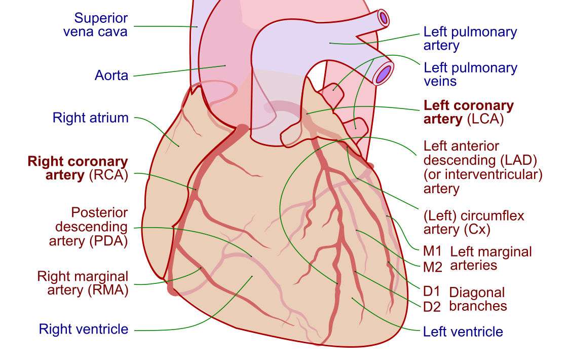
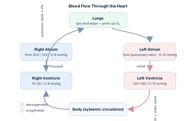
If a right heart study is added, a balloon-tipped catheter records pressures through the right atrium, right ventricle, pulmonary artery, and wedge positions — the data behind measurements like cardiac output and cardiac index. This is where a firm grasp of hemodynamic principles turns raw waveforms into a diagnosis. Throughout, you are awake but relaxed under light sedation — most people feel pressure but little pain, and the whole thing is often over before they expect.
When it becomes an intervention (and takes longer)
Here is where the timeline can change on the spot. If the diagnostic angiogram reveals a tight, flow-limiting blockage, the cardiologist may move directly into treatment in the same session — a strategy called ad hoc percutaneous coronary intervention. If you want the full contrast between the two, our explainer on PCI vs. cardiac catheterization breaks it down, but the timing headline is simple: intervention adds time.
A treatment adds several steps to the diagnostic study. The patient receives blood-thinning medication, a hair-thin guidewire is steered across the blockage, a balloon is inflated to open the vessel (angioplasty), and a stent — usually a drug-eluting stent — is deployed to hold the artery open. Each step takes time, and difficult anatomy can add much more.
Because a stent has now been placed, recovery is longer too. Blood thinners must stay active, the access site needs more careful monitoring, and observation may extend for several hours or overnight. So a case that would have been a tidy 40-minute diagnostic study can reasonably become a 90-minute-plus procedure with a much longer tail — an entirely normal and often welcome outcome, since the problem was found and fixed in one trip.
Recovery time: the part people underestimate
The recovery phase is where the wrist-versus-groin decision earns its keep, and it is usually the longest single block of your day. The reason is straightforward: the sheath went into an artery, and once it comes out, that artery has to seal so it does not bleed.
With a radial (wrist) approach, a small compression band is placed and you can typically sit up and even walk soon after. Recovery observation often runs about two to three hours, and many patients go home the same day. The wrist approach has become the preferred default in many labs precisely because it is more comfortable and gets people moving faster, with less bleeding risk.
With a femoral (groin) approach, the artery is larger and higher-pressure, so you must lie flat and keep the leg still for several hours — commonly three to six — while the puncture seals, whether by manual pressure or a closure device. That immobility, not the procedure, is what makes the groin route feel longer.
| Feature | Radial (wrist) | Femoral (groin) |
|---|---|---|
| Lie-flat time | None to minimal | Several hours |
| Typical recovery watch | 2–3 hours | 3–6 hours |
| Bleeding risk | Lower | Higher |
| Same-day discharge | Very common | Common, but slower |
During recovery, nurses check the access site repeatedly, monitor your rhythm and blood pressure, and encourage fluids to help flush the contrast dye through your kidneys. If you had only a diagnostic study, you are usually cleared to go home once you are stable and the site is secure.
Total time, start to finish: three scenarios
Pulling the pieces together, it helps to picture a few realistic days. These are illustrative ranges, not guarantees — your team's routine and your specific situation set the true numbers.
- Routine diagnostic, radial access. Arrive, prep for about an hour, a 30-minute procedure, then two to three hours of recovery. Total: roughly 4 to 5 hours, home the same day.
- Diagnostic that becomes a stent (ad hoc PCI). Same prep, but the procedure runs an hour or more, and recovery extends to four-plus hours or overnight. Total: 6 hours to an overnight stay.
- Emergency heart-attack (primary PCI). A different animal entirely. The team works to open the blocked artery as fast as possible — the door-to-balloon clock is measured in minutes — followed by admission to a cardiac unit for close monitoring. Total hospital time is measured in days, not hours.
The emergency scenario is a reminder of why speed and skill in the lab matter. When a patient arrives with an ongoing heart attack, a confident read of the STEMI ECG triggers a race to reopen the artery, because in that setting time is heart muscle. The same catheter that leisurely maps arteries in an elective study becomes a life-saving tool moving against the clock.
What you can (and can't) control about the timing
Patients often ask whether they can do anything to make the day go faster. Mostly the timeline is set by biology and by the procedure, but a few things genuinely help — and a few common worries are out of your hands.
Within your influence:
- Follow fasting and medication instructions exactly. A missed instruction can delay or postpone the case.
- Arrive on time with your medication list. Prep goes faster when the team is not chasing information.
- Stay hydrated as directed and stay still during recovery. Moving the leg too soon after a femoral case can restart bleeding and reset the clock.
- Ask questions up front. Understanding the plan reduces anxiety, which can lower the sedation and monitoring you need.
Outside your control: the complexity of your arteries, whether a blockage needs treating, how your body responds to sedation, and — realistically — the lab's schedule that day. Emergencies always take priority, and an unexpected STEMI can bump an elective case. That is not a failure of planning; it is the system working exactly as it should, since those cases cannot wait.
Who keeps the procedure on track
A cath case runs smoothly because of a coordinated team, and the pacing you experience is largely their doing. The cardiologist directs the case, but a registered cardiovascular invasive specialist (RCIS) and other cardiovascular technologists keep it moving — scrubbing in, managing the hemodynamic monitoring, running the imaging and physiology equipment, and preparing devices before they are needed.
Much of what makes a case efficient is invisible to the patient: anticipating the next catheter, having the right balloon or stent ready, catching a subtle rhythm change on the monitor before it becomes a problem. That real-time vigilance draws on deep command of cardiac anatomy and waveform interpretation — the very skills the RCIS credential certifies. A well-drilled team is why the procedure so often finishes faster than patients feared.
Safety, not speed, is always the governing principle. If the team slows down to double-check an access site, confirm a pressure measurement, or manage a blip in your rhythm, that pause is time well spent. The goal is a complete, safe study — and, when needed, a complete, safe treatment — not a fast one.
Key takeaways
- A diagnostic cardiac catheterization usually takes about 20–45 minutes of procedure time, but the full day runs roughly 4–6 hours once prep and recovery are counted.
- The biggest driver of duration is whether the study stays diagnostic or becomes an intervention — a stent adds procedure time and lengthens recovery, sometimes to an overnight stay.
- Prep (60–90 minutes) covers blood work, IV, medication review, and monitoring — time that prevents complications rather than wasting them.
- Recovery is usually the longest block: a radial (wrist) approach lets you move sooner (2–3 hours), while a femoral (groin) approach requires lying flat for several hours.
- An emergency primary PCI for a heart attack is a different timeline entirely — fast on the table, then days of monitoring, because time is heart muscle.
- You can help by following fasting and medication instructions, arriving prepared, staying still during recovery, and bringing a driver.
- A skilled cath-lab team keeps the case efficient and safe; a deliberate pause is a feature, not a delay.
- This article is educational and not medical advice — your own cardiologist and care team set the real expectations for your procedure.
Curious what happens in the cath lab?
See who is in the room and what the equipment does.
What Is a Cardiac Cath Lab →Frequently asked questions
How long does a cardiac catheterization take?
A diagnostic cardiac catheterization usually takes about 20 to 45 minutes of actual procedure time. However, when you add check-in, prep, sedation, and recovery, plan on being at the hospital for roughly 4 to 6 hours. If the study turns into a treatment such as a stent, both the procedure and the recovery take longer.
Why do I have to stay for hours if the procedure is only 30 minutes?
Because most of your day is prep and recovery, not the procedure. Prep involves blood work, an IV, a medication review, and monitoring, which takes about an hour or more. Afterward, the artery used for access has to seal safely, and you are watched for bleeding and rhythm changes for two to six hours depending on the access site.
Does a cardiac cath take longer if I need a stent?
Yes. Placing a stent adds several steps to the diagnostic study — blood thinners, crossing the blockage with a guidewire, balloon angioplasty, and deploying the stent. That commonly turns a 30- to 40-minute diagnostic case into a 45- to 90-minute or longer procedure, and the recovery period also lengthens, sometimes to an overnight stay.
How long is recovery after a cardiac catheterization?
Recovery depends on the access site. With a wrist (radial) approach, a compression band is used and observation usually runs two to three hours, with same-day discharge common. With a groin (femoral) approach, you must lie flat and keep the leg still for about three to six hours while the larger artery seals, so recovery feels longer.
Is a wrist or groin approach faster to recover from?
The wrist (radial) approach is generally faster and more comfortable to recover from, because the smaller artery seals with a compression band and you can sit up and walk sooner. The groin (femoral) approach requires lying flat for several hours, so it typically means a longer recovery watch and a higher bleeding risk. Many labs now prefer the wrist route for these reasons.
How long does an emergency cardiac cath for a heart attack take?
An emergency catheterization with primary PCI is deliberately fast on the table — the team works to reopen the blocked artery within minutes, tracked by the door-to-balloon time. But total hospital time is much longer, usually several days, because a heart attack requires admission to a cardiac unit for close monitoring and further treatment.
How long before a cardiac cath should I stop eating?
You are typically asked not to eat or drink for several hours before the procedure, often from midnight the night before or for at least six to eight hours prior, though clear liquids may sometimes be allowed. Your care team gives the exact instructions, which also cover which medications to hold or take, so follow their guidance precisely to avoid delaying the case.
Can I drive home after a cardiac catheterization?
No. Because you receive sedation and an artery has been accessed, you should not drive for the rest of the day. Arrange for someone to drive you home. Even after a quick diagnostic study, most centers require a responsible adult to take you home and, ideally, stay with you for the first evening.
Will I be awake during the cardiac catheterization?
Usually yes. Most cardiac catheterizations are done under light sedation, so you are awake but relaxed and drowsy rather than fully asleep. A local anesthetic numbs the access site, and while you may feel pressure or brief warmth from the contrast dye, the procedure is generally not painful. You can often talk with the team during the study.
Sources & further reading
- Cardiovascular Credentialing International (CCI)
- American College of Cardiology
- American Heart Association
- MedlinePlus (U.S. National Library of Medicine)
External links are provided for reference; always confirm current details with the official source.