Cardiac Index: Normal Range, Formula & Clinical Meaning
Cardiac index is cardiac output tailored to body size — the single number that lets you compare a 110-pound patient with a 250-pound one and know whose heart is actually keeping up. This guide covers the formula, the normal range, how it differs from cardiac output, and what a low index is really telling you.
What is cardiac index?
Cardiac index (CI) is cardiac output normalized to a patient's body surface area. In plain terms, it takes the total volume of blood the heart pumps each minute and divides it by how big the person is, giving a per-square-meter value that means the same thing whether the patient is petite or large.
Why bother? Because raw cardiac output is misleading on its own. A cardiac output of 4 liters per minute might be perfectly healthy for a small adult but dangerously inadequate for a tall, muscular one — their tissues simply demand more blood. By indexing to body size, cardiac index answers the question that actually matters at the bedside: is this heart delivering enough flow for this particular body?
You will meet cardiac index constantly in the cath lab, the ICU, and on the RCIS exam. It sits at the heart of hemodynamic assessment, and understanding it is the gateway to reading everything else — from systemic vascular resistance to the pressure tracings you interpret during a right-heart catheterization. If you want the broader picture, our hemodynamics study guide ties all of these numbers together.
This article is educational and not medical advice. Clinical thresholds vary by patient and institution; always follow local protocols and current guidelines.
Cardiac index formula
The formula is refreshingly simple:
Cardiac Index = Cardiac Output ÷ Body Surface Area
CI (L/min/m²) = CO (L/min) ÷ BSA (m²)
Every part of that equation is worth unpacking:
- Cardiac output (CO) is the volume of blood the heart ejects per minute, measured in liters per minute. It equals stroke volume multiplied by heart rate. In the cath lab, CO is usually obtained by thermodilution or the Fick method — see our cardiac output guide for how each technique works.
- Body surface area (BSA) is an estimate of a person's total skin area in square meters, calculated from height and weight. Most labs use the Du Bois or Mosteller equation. A typical adult BSA is roughly 1.6 to 2.0 m².
So if a patient has a cardiac output of 5.0 L/min and a body surface area of 1.9 m², their cardiac index is 5.0 ÷ 1.9 ≈ 2.63 L/min/m² — comfortably normal.
Because BSA is almost always larger than 1.0, cardiac index is always a smaller number than cardiac output. That relationship is a handy sanity check: if someone quotes a "cardiac index" of 5 L/min/m², they have almost certainly confused it with cardiac output.
What is a normal cardiac index?
A normal cardiac index in a resting adult is approximately 2.5 to 4.0 L/min/m². Some references cite a slightly wider band of 2.2 to 4.2, and the exact cutoffs vary by textbook and monitoring device, so treat these numbers as clinical guideposts rather than hard lines.
| Cardiac index (L/min/m²) | Interpretation |
|---|---|
| > 4.0 | High output — sepsis, thyrotoxicosis, anemia, arteriovenous shunt, pregnancy |
| 2.5 – 4.0 | Normal resting range |
| 2.2 – 2.5 | Low-normal / early impairment; watch closely |
| 1.8 – 2.2 | Reduced output; often symptomatic heart failure |
| < 1.8 | Cardiogenic shock territory (especially with signs of hypoperfusion) |
Two thresholds are worth memorizing because they show up on exams and in real decision-making. A cardiac index below about 2.2 L/min/m² is a classic marker of significant cardiac impairment, and a value under 1.8 L/min/m² with evidence of poor perfusion defines cardiogenic shock in the traditional Forrester classification. Above the top of the range, a high cardiac index usually reflects a body demanding extra flow — as in sepsis or hyperthyroidism — rather than a heart that is simply overperforming.
Keep in mind that cardiac index falls naturally with age as resting metabolic demand declines, and it rises with exercise, fever, and pregnancy. A single value is a snapshot; the trend over time often tells you more than any one reading.
Cardiac index vs cardiac output
This is the distinction students trip over most, so let us be precise. Cardiac output is the absolute volume of blood pumped per minute; cardiac index is that same output divided by body surface area. Cardiac index is simply cardiac output made comparable across people of different sizes.
| Cardiac output (CO) | Cardiac index (CI) | |
|---|---|---|
| What it measures | Total blood flow per minute | Blood flow per minute, per m² of body |
| Units | L/min | L/min/m² |
| Normal range | ~4 – 8 L/min | ~2.5 – 4.0 L/min/m² |
| Adjusts for body size? | No | Yes |
| Best for | A single patient over time | Comparing patients / defining shock |
An analogy helps. Cardiac output is like a car's total horsepower; cardiac index is horsepower-per-ton. A big truck and a small hatchback can have the same horsepower, but the number that predicts how briskly each actually moves is the power-to-weight ratio. Cardiac index is the cardiovascular power-to-weight ratio.
The practical payoff: two patients can share an identical cardiac output of 4.5 L/min, yet if one has a BSA of 1.5 m² and the other 2.4 m², their cardiac indices are 3.0 versus 1.9 L/min/m². The first patient is fine; the second may be sliding toward shock. That is exactly why intensivists prefer the index when they classify shock states. We break the comparison down further in our dedicated cardiac output explainer.
How cardiac index is measured
You cannot measure cardiac index directly — it is always calculated from a measured cardiac output and a computed body surface area. Getting the cardiac output right is therefore the whole game, and there are several established methods.
- Thermodilution is the workhorse of the cath lab and ICU. A bolus of cold saline is injected through a Swan-Ganz (pulmonary artery) catheter, and a thermistor downstream measures how quickly the blood warms back up. The area under the temperature curve is inversely proportional to flow. Modern PA catheters can also give continuous cardiac output.
- The Fick principle derives cardiac output from oxygen consumption divided by the arteriovenous oxygen difference. It is the reference standard in low-output states where thermodilution can be unreliable. Our Fick calculator walks through the arithmetic.
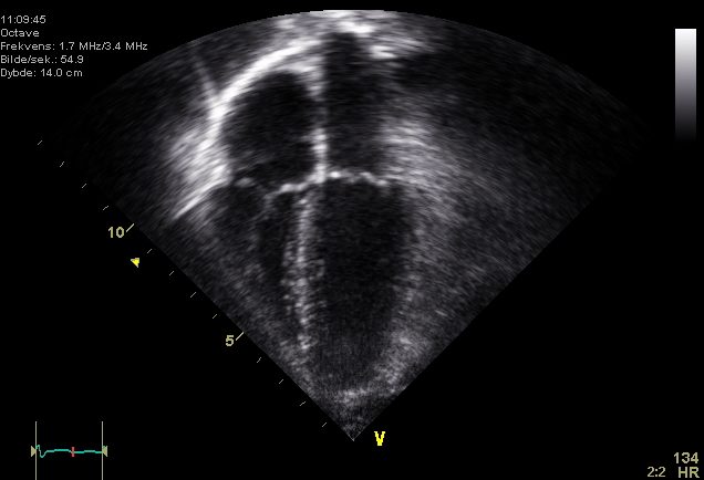
- Echocardiography estimates stroke volume from the left ventricular outflow tract and multiplies by heart rate — a noninvasive option covered in our echocardiogram guide. The ejection fraction often travels alongside these measurements.
- Pulse-contour and bioreactance devices analyze the arterial waveform or thoracic electrical signals to track cardiac output continuously and less invasively.
Whichever method supplies the output, the software or the operator then divides by the patient's BSA to report the index. Because errors in BSA propagate directly into the index, an inaccurate height or weight entry can quietly skew every derived number.
What does a low cardiac index mean?
A low cardiac index means the heart is not delivering enough blood for the body's size and needs — the hemodynamic signature of a failing pump. As the index drops, tissues receive less oxygen, and the body responds with compensatory changes you can often see at the bedside before the numbers even print.
Common causes of a low cardiac index include:
- Cardiogenic shock after a large myocardial infarction, where damaged muscle can no longer pump effectively
- Advanced heart failure and cardiomyopathy
- Severe valvular disease, such as critical aortic stenosis (see our aortic valve area guide)
- Cardiac tamponade, where fluid around the heart restricts filling
- Massive pulmonary embolism straining the right ventricle
- Profound bradyarrhythmias or poorly tolerated tachyarrhythmias
Clinically, a genuinely low index produces cool, mottled extremities, weak pulses, a narrow pulse pressure, rising lactate, falling urine output, and altered mental status. When the index sinks below 2.2 L/min/m² and especially below 1.8, clinicians escalate quickly — inotropes, vasopressors, and mechanical support such as an intra-aortic balloon pump all aim to lift that number back into a survivable range. The index is one of the vital signs that guides how aggressive that support needs to be.
What does a high cardiac index mean?
A high cardiac index — above roughly 4.0 L/min/m² — sounds like a good thing but usually is not. It typically means the body is demanding more flow, and the heart is being driven hard to supply it. The pump may be healthy, but something is forcing it to overwork.
High-output states include:
- Sepsis and early septic shock, where widespread vasodilation drops resistance and the heart compensates with a high output. This is the classic "warm shock" — high index, low systemic vascular resistance.
- Thyrotoxicosis, which cranks up metabolic rate and heart rate.
- Severe anemia, where thin blood must circulate faster to deliver the same oxygen.
- Large arteriovenous shunts or fistulas, including dialysis access.
- Pregnancy, beriberi (thiamine deficiency), and Paget disease.
The key teaching point is that a high cardiac index is rarely reassuring on its own. In sepsis, a high index alongside a low resistance and a rising lactate signals distributive shock — the flow is high but it is being distributed inefficiently, and tissues can still starve. Reading the index together with resistance and filling pressures is what separates the useful clinician from someone staring at a single number.
Cardiac index in the cath lab and on the RCIS exam
For cardiovascular technologists and RCIS candidates, cardiac index is not an abstraction — it is something you help generate during nearly every right-heart catheterization. When you run a thermodilution set or perform an oximetry run for a Fick calculation, the resulting cardiac index feeds directly into the physician's assessment of pump function, shunt severity, and readiness for interventions.
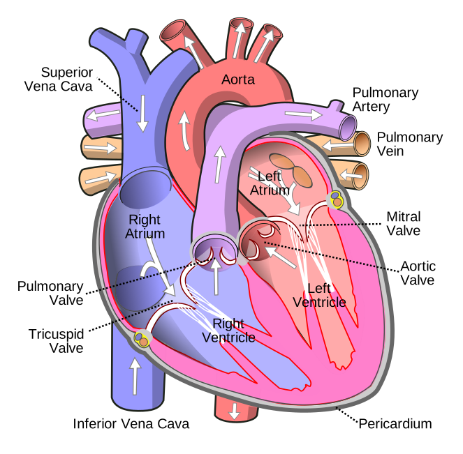
The exam loves to test whether you can move fluently between related values. Expect questions that give you a cardiac output and BSA and ask for the index, or that hand you an index and ask you to classify the patient. You should also be ready to distinguish the index from cardiac output, stroke volume, and resistance in a single scenario. Brush up on the surrounding anatomy in our cardiac anatomy guide, then drill the numbers with our hemodynamics practice questions.
Because the index depends on an accurate output, the same measurement pitfalls apply: shivering or arrhythmia during thermodilution, incorrect injectate temperature, or a wedged catheter can all throw the number off. Recognizing an implausible index — and knowing to repeat the measurement — is a mark of a skilled tech.
Key takeaways
- Cardiac index = cardiac output ÷ body surface area, expressed in L/min/m². It scales blood flow to body size.
- A normal resting cardiac index is about 2.5 to 4.0 L/min/m²; below 2.2 signals meaningful impairment and below 1.8 with hypoperfusion defines cardiogenic shock.
- Cardiac index vs cardiac output: output is total flow (L/min); index is flow adjusted for size (L/min/m²). The index is better for comparing patients and defining shock.
- A low index points to pump failure — MI, heart failure, tamponade, severe valve disease — and drives escalation to inotropes and mechanical support.
- A high index usually reflects high demand — sepsis, thyrotoxicosis, anemia — and is rarely reassuring on its own.
- Cardiac index is calculated, not measured directly; accuracy depends on a reliable cardiac output and a correct body surface area.
- This content is educational, not medical advice, and clinical thresholds are evolving — always defer to current guidelines and local protocols.
Calculate cardiac index
Enter cardiac output and body surface area to get cardiac index with interpretation.
Open the Cardiac Index Calculator →Frequently asked questions
What is a normal cardiac index?
In a resting adult, a normal cardiac index is roughly 2.5 to 4.0 L/min/m², with some references citing 2.2 to 4.2. Values fall naturally with age and rise with exercise, fever, and pregnancy, so interpret any single number in clinical context.
What is the cardiac index formula?
Cardiac index equals cardiac output divided by body surface area: CI (L/min/m²) = CO (L/min) ÷ BSA (m²). Cardiac output itself is stroke volume times heart rate, and BSA is estimated from height and weight.
What is the difference between cardiac index and cardiac output?
Cardiac output is the total volume of blood pumped per minute (L/min). Cardiac index is that output divided by body surface area (L/min/m²), which lets you compare patients of different sizes. The index is preferred for defining and classifying shock.
What does a low cardiac index mean?
A low cardiac index means the heart is not delivering enough blood for the body's needs — a sign of a failing pump. Causes include myocardial infarction, heart failure, cardiac tamponade, and severe valve disease. Below 1.8 L/min/m² with poor perfusion suggests cardiogenic shock.
What is a dangerously low cardiac index?
A cardiac index below about 2.2 L/min/m² indicates significant cardiac impairment, and a value under 1.8 L/min/m² accompanied by signs of hypoperfusion (cool skin, low urine output, rising lactate) is in the cardiogenic-shock range and typically prompts urgent treatment.
Why is cardiac index used instead of cardiac output?
Because raw cardiac output ignores body size. A 4 L/min output is normal for a small adult but inadequate for a large one. Indexing to body surface area makes the value comparable across patients and lets clinicians apply consistent thresholds.
What causes a high cardiac index?
High cardiac index reflects increased demand rather than a stronger heart. Common causes are sepsis, thyrotoxicosis, severe anemia, arteriovenous shunts, and pregnancy. In sepsis it pairs with low systemic vascular resistance — the pattern of distributive shock.
How is cardiac index measured?
It is calculated, not measured directly. Cardiac output is obtained by thermodilution through a Swan-Ganz catheter, by the Fick oxygen method, or by echocardiography, then divided by the patient's body surface area to give the index.
Is cardiac index the same as ejection fraction?
No. Cardiac index measures blood flow per minute scaled to body size (L/min/m²), while ejection fraction is the percentage of blood ejected from the left ventricle with each beat. Both assess cardiac function but describe different things.
Sources & further reading
- Cardiovascular Credentialing International (CCI)
- American College of Cardiology
- American Heart Association
- MedlinePlus (U.S. National Library of Medicine)
External links are provided for reference; always confirm current details with the official source.