Cardiac Tamponade Hemodynamics
Cardiac tamponade is what happens when fluid piles up in the pericardial sac faster than the heart can adapt — squeezing every chamber until filling fails and output collapses. This guide breaks down the hemodynamics: equalized pressures, the blunted y descent, pulsus paradoxus, and how tamponade differs from constrictive pericarditis on the tracing.
- What cardiac tamponade actually is
- Cardiac tamponade hemodynamics in one picture
- Equalization of diastolic pressures
- The blunted y descent on the RA waveform
- Pulsus paradoxus explained
- Tamponade vs constriction on the tracing
- Recognizing tamponade at the bedside
- Iatrogenic tamponade in the cath lab
- Why tamponade physiology matters for the RCIS exam
- Key takeaways
What cardiac tamponade actually is
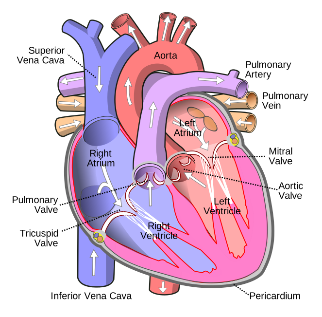
Cardiac tamponade is a state in which fluid accumulating inside the pericardial sac raises intrapericardial pressure enough to compress the heart and choke off its filling. The pericardium is a tough, relatively stiff bag surrounding the heart. It normally holds only a few milliliters of lubricating fluid. When blood, pus, or an effusion collects in that space, pressure inside the sac climbs — and because the heart lives inside the same sac, that external pressure is transmitted straight onto the cardiac chambers.
The key idea is not how much fluid there is, but how fast it arrives. A slowly growing chronic effusion can stretch to a liter or more without causing tamponade because the pericardium has time to accommodate. A brisk bleed after a coronary perforation or a stab wound can cause life-threatening tamponade with as little as 150–200 mL, because the sac cannot stretch quickly enough. That is the pericardial compliance curve at work: gentle at first, then brutally steep once the reserve volume is used up.
Understanding tamponade means understanding pressure and flow together, so if you want the broader framework the pressures live in, our hemodynamics study guide ties these tracings back to the chambers that generate them.
This article is educational and not medical advice. Tamponade is a clinical emergency judged at the bedside with echocardiography and the full picture — always follow current guidelines and local protocols.
Cardiac tamponade hemodynamics in one picture
The whole story of cardiac tamponade hemodynamics flows from a single mechanical problem: the pericardial pressure is now high enough to compete with the pressures inside the heart's chambers. Filling a chamber requires that the pressure inside it exceed the pressure squeezing it from outside. Once intrapericardial pressure rises to meet — and then match — the diastolic pressures of the right atrium and right ventricle, those thin-walled right-heart chambers can barely fill.
Three things follow directly from that:
- Rising, then equalized, filling pressures. Right atrial, right ventricular diastolic, pulmonary artery diastolic, and pulmonary capillary wedge pressures all climb and converge toward the same elevated value.
- Impaired filling, so falling stroke volume. If chambers cannot fill, they cannot eject a normal volume. Stroke volume drops, and the body compensates with tachycardia and vasoconstriction to defend blood pressure.
- Exaggerated interventricular dependence. Because total cardiac volume is fixed by the tense sac, the two ventricles compete for space through the septum — the mechanism behind pulsus paradoxus, covered below.
The compensatory tachycardia and clamped-down systemic vascular resistance are the body's attempt to hold mean arterial pressure while stroke volume falls. You can see how that fits the larger compensation picture in our overview of shock hemodynamics and how afterload is quantified in the piece on systemic vascular resistance. When those compensations exhaust, obstructive shock and pulseless electrical activity follow fast.
Equalization of diastolic pressures
The single most tested hemodynamic hallmark of tamponade is equalization — the near-identical elevation of the diastolic filling pressures across the right and left heart. On a right-heart catheterization with a Swan-Ganz catheter, you see the right atrial mean pressure, the right ventricular end-diastolic pressure, the pulmonary artery diastolic pressure, and the pulmonary capillary wedge pressure all cluster within a few mmHg of one another, usually somewhere in the 15–20 mmHg range.
Why do they converge? Because the pericardial pressure has become the dominant force setting diastolic pressure in every chamber. Instead of each chamber having its own filling pressure, they are all effectively reporting the same thing: the pressure inside the sac squeezing them. The heart's chambers stop behaving independently and start behaving like one compressed unit.
| Pressure | Normal (mmHg) | In tamponade (mmHg) |
|---|---|---|
| Right atrial (mean) | 2–6 | 15–20 |
| RV end-diastolic | 2–8 | 15–20 |
| PA diastolic | 8–15 | 15–20 |
| PCWP (wedge) | 6–12 | 15–20 |
Notice how the normal values span a wide spread — the right atrium normally sits far below the wedge. In tamponade they collapse onto one number. That convergence, often written as equalization of diastolic pressures, is the sentence graders love. A useful RCIS shorthand is that within roughly 5 mmHg of each other counts as equalized.
The blunted y descent on the RA waveform
To read the next clue you need the normal right atrial pressure tracing, which has two downward deflections: the x descent (during ventricular systole, as the atrium relaxes and the tricuspid annulus pulls down) and the y descent (in early diastole, when the tricuspid valve opens and the atrium empties rapidly into the ventricle).
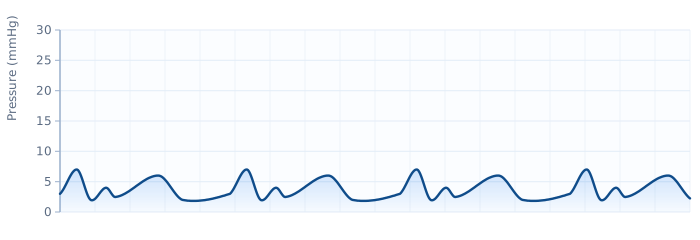
In tamponade, the y descent is blunted or absent. Here is the logic: the y descent depends on blood rushing out of the atrium into a ventricle that has room to receive it. But in tamponade the ventricle is being crushed from outside all through diastole — it has no room. When the tricuspid valve opens, blood cannot pour in, so the atrial pressure does not fall the way it should. The result is a tracing where the x descent is preserved (systole still transiently decompresses the atrium a little) but the y descent flattens out.
This detail matters because it separates tamponade from its main mimic. In constrictive pericarditis, early diastolic filling is actually rapid — it just stops abruptly — so the y descent is preserved and often exaggerated. Tamponade blunts the y descent; constriction keeps it. Hold onto that contrast; it is the crux of the comparison section below.
Pulsus paradoxus explained
Pulsus paradoxus is an exaggerated fall in systolic blood pressure during inspiration — classically defined as a drop of more than 10 mmHg with a normal quiet breath. It is one of the most reliable physical-exam signs of significant tamponade, and it confuses students until the mechanism clicks.
Start with normal physiology. When you breathe in, the drop in intrathoracic pressure pulls extra blood into the right heart, so right-sided filling rises with inspiration. In a healthy chest, the two ventricles have room to accommodate this and left-sided output barely changes. Now add a tense pericardial sac that fixes the total volume the heart is allowed to occupy. On inspiration, the extra blood filling the right ventricle has nowhere to go but to bow the interventricular septum leftward, into the left ventricle. That intrudes on the left ventricular cavity, reduces its filling, drops its stroke volume, and so drops systolic pressure with each breath in. That is ventricular interdependence made visible at the wrist and on the arterial line.
It is called "paradoxus" for a historical reason: Kussmaul noticed the pulse could disappear at the wrist during inspiration even while the heartbeat was still audible — the pulse seemed to vanish paradoxically. It is not truly paradoxical; it is just an exaggeration of a normal inspiratory dip.
| Feature | Detail |
|---|---|
| Definition | Inspiratory systolic BP fall > 10 mmHg |
| Mechanism | Exaggerated ventricular interdependence in a fixed pericardial volume |
| How to measure | Slowly deflate a BP cuff; note the gap between the first Korotkoff sounds (heard only in expiration) and when they are heard throughout the cycle |
| Also seen in | Severe asthma/COPD, massive pulmonary embolism, tension pneumothorax, hypovolemic shock |
Because pulsus paradoxus also appears in severe airway disease and large pulmonary embolism, it supports the diagnosis but does not clinch it — echocardiography does. And a few tamponade situations mute it: coexisting aortic regurgitation, a large atrial septal defect, or severe left ventricular dysfunction can blunt the sign even when tamponade is present.
Tamponade vs constriction on the tracing
The most exam-relevant comparison — and a genuinely tricky bedside call — is tamponade vs constriction (constrictive pericarditis). Both squeeze the heart, both raise and equalize filling pressures, and both can cause right heart failure signs. The difference lies in timing: tamponade compresses the heart throughout the whole cardiac cycle, whereas constriction lets the heart fill briefly and freely in very early diastole before a rigid pericardial shell abruptly stops it.
That single mechanical distinction drives every waveform difference below.
| Feature | Cardiac tamponade | Constrictive pericarditis |
|---|---|---|
| Compression | Throughout diastole | Only after early rapid filling |
| y descent (RA) | Blunted / absent | Preserved, steep (prominent) |
| RV/LV waveform | No dip-plateau | Square-root / dip-and-plateau sign |
| Equalization of diastolic pressures | Yes | Yes |
| Pulsus paradoxus | Common, prominent | Often absent or mild |
| Kussmaul sign (JVP rises with inspiration) | Usually absent | Often present |
| Ventricular interdependence | Present | Present (discordant RV/LV pressures) |
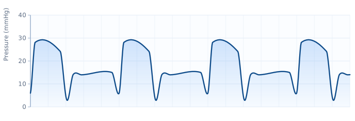
Two waveform features do the heavy lifting. First, the y descent: blunted in tamponade, sharp and preserved in constriction — a direct readout of whether early filling is blocked or brief-but-free. Second, the square-root sign (dip-and-plateau) on the ventricular tracing: present in constriction, absent in tamponade. If you see a crisp square-root sign with a steep y descent, think constriction. If you see equalized pressures with a flat y descent and pulsus paradoxus, think tamponade.
Recognizing tamponade at the bedside
Hemodynamic tracings are the exam's focus, but real tamponade is often recognized clinically first, then confirmed with imaging. The classic teaching triad is Beck's triad: hypotension, muffled heart sounds, and distended neck veins (elevated jugular venous pressure). In practice the full triad appears in a minority of patients, so waiting for all three is a mistake.
More sensitive and more common findings include:
- Tachycardia — the earliest compensation for a falling stroke volume.
- Pulsus paradoxus greater than 10 mmHg, as described above.
- Elevated JVP with a blunted y descent visible in the neck veins.
- Dyspnea and a sense of air hunger, often with the patient sitting forward.
- Low ECG voltage and electrical alternans — the QRS complexes swing in amplitude beat to beat as the heart swings in the fluid. If you want to sharpen waveform reading in general, our ECG interpretation guide and the hands-on ECG strip practice set build that pattern-recognition muscle.
Echocardiography is the confirmatory test. It shows the effusion, right atrial systolic collapse, right ventricular early-diastolic collapse, a plethoric inferior vena cava that does not collapse with inspiration, and exaggerated respiratory variation in transvalvular inflow — the echo equivalent of pulsus paradoxus. Our overview of the echocardiogram covers how those views are obtained. Because RA and RV collapse can precede a big drop in blood pressure, echo frequently catches tamponade physiology before the classic vitals declare themselves.
Iatrogenic tamponade in the cath lab
For RCIS candidates and cath-lab staff, tamponade is not an abstract cardiology-textbook entity — it is a procedural complication you may see develop in real time. Coronary or cardiac perforation, guidewire exit, lead placement during pacing, and structural interventions can all bleed into the pericardium. When they do, the tamponade is usually acute, so even a small, fast accumulation can be catastrophic.
Warning signs on the table: unexplained hypotension, a rising heart rate, a new drop in the arterial waveform, a patient reporting chest pressure, and on fluoroscopy a widening cardiac silhouette with a sluggish or motionless heart border. Confirming rests on rapid echocardiography — a skill set that overlaps with the work of the cardiovascular ultrasound technologist. Understanding what a normal case looks like helps you spot the abnormal one, and our primer on what a cardiac cath lab is sets that scene.
The definitive treatment is drainage — pericardiocentesis (needle aspiration under echo or fluoroscopic guidance) or a surgical pericardial window for recurrent, loculated, or clotted collections. Removing even a modest volume of fluid can drop intrapericardial pressure below the steep part of the compliance curve and restore filling dramatically, which is why patients can improve within seconds of a successful tap. Intravenous fluids and inotropes are only a bridge; they buy minutes, not a cure.
Why tamponade physiology matters for the RCIS exam
Tamponade is a favorite RCIS topic because it forces you to reason across pressures, waveforms, and clinical signs at once — exactly the integrated thinking the credential is testing. A single well-written question can hinge on recognizing equalized diastolic pressures, then choosing the blunted y descent (not the preserved one) as the distinguishing feature from constriction.
It also sits at the intersection of several core concepts you will study elsewhere: the falling stroke volume that drives compensatory tachycardia, the cardiac output that ultimately falls when compensation fails, and the pressures a Swan-Ganz catheter reports. When you can trace how one stiff sac of fluid ripples through all of those numbers, you have internalized the physiology rather than memorized a list.
Practice cements it. Working through the hemodynamics question bank alongside the cardiac anatomy guide will keep the tamponade pattern fresh, and mixing in waveform identification builds the reflex of naming a tracing at a glance — the single most valuable skill on the hemodynamics portion of the test.
Key takeaways
- Tamponade compresses the heart throughout diastole — rate of fluid accumulation matters more than absolute volume.
- Equalization of diastolic pressures (RA ≈ RVEDP ≈ PA diastolic ≈ PCWP, often 15–20 mmHg) is the hemodynamic hallmark.
- The y descent is blunted or absent in tamponade because the ventricle cannot accept early-diastolic inflow — the opposite of constriction's steep, preserved y descent.
- Pulsus paradoxus (inspiratory systolic BP fall > 10 mmHg) reflects exaggerated ventricular interdependence inside a fixed pericardial volume.
- Tamponade vs constriction: both equalize pressures, but constriction shows a preserved y descent and a square-root (dip-and-plateau) ventricular sign that tamponade lacks.
- Echocardiography confirms it; drainage (pericardiocentesis or surgical window) treats it — fluids and inotropes only bridge.
- This article is educational and not medical advice; always follow current guidelines and your local protocols.
Practise tamponade & waveforms
Test hemodynamic pattern-recognition with explanations.
Practise Hemodynamics →Frequently asked questions
What is the classic hemodynamic hallmark of cardiac tamponade?
Equalization of the diastolic filling pressures. In tamponade the right atrial mean pressure, right ventricular end-diastolic pressure, pulmonary artery diastolic pressure, and pulmonary capillary wedge pressure all rise and converge toward the same elevated value, typically in the 15–20 mmHg range, because the high intrapericardial pressure dominates diastolic filling in every chamber.
Why is the y descent blunted in cardiac tamponade?
The y descent normally reflects rapid early-diastolic emptying of the atrium into the ventricle when the AV valve opens. In tamponade the ventricle is compressed throughout diastole and has no room to accept that inflow, so atrial pressure does not fall when the valve opens. The result is a blunted or absent y descent, with the x descent relatively preserved.
How does tamponade differ from constrictive pericarditis on hemodynamics?
Both equalize diastolic pressures, but the y descent and ventricular waveform separate them. Tamponade compresses the heart all through diastole, giving a blunted y descent and no dip-plateau. Constriction allows brief rapid early filling that stops abruptly, producing a preserved, steep y descent and a square-root (dip-and-plateau) sign on the ventricular tracing. Pulsus paradoxus is prominent in tamponade and often absent in constriction, while the Kussmaul sign is more typical of constriction.
What is pulsus paradoxus and why does it occur in tamponade?
Pulsus paradoxus is an exaggerated fall in systolic blood pressure of more than 10 mmHg during inspiration. In a tense pericardial sac the total cardiac volume is fixed, so inspiratory increases in right heart filling push the interventricular septum toward the left ventricle, reducing its filling and stroke volume with each breath in. It is a bedside marker of ventricular interdependence.
How much fluid does it take to cause cardiac tamponade?
There is no fixed number — the rate of accumulation matters more than the amount. A rapidly bleeding effusion can cause tamponade with only 150–200 mL because the pericardium cannot stretch fast enough, while a slowly developing chronic effusion may reach a liter or more without tamponade because the sac has time to accommodate.
Is pulsus paradoxus always present in tamponade?
No. It is common and supportive but not universal. Coexisting aortic regurgitation, a large atrial septal defect, severe left ventricular dysfunction, or low-pressure and regional tamponade can mute or eliminate the sign even when tamponade is hemodynamically significant. Its absence does not rule tamponade out, so echocardiography remains the deciding test.
What is Beck's triad?
Beck's triad is the classic clinical trio of acute cardiac tamponade: hypotension, muffled or distant heart sounds, and distended neck veins from elevated jugular venous pressure. The full triad is present in only a minority of patients, so its absence should never be used to exclude tamponade.
How is cardiac tamponade diagnosed and treated?
Diagnosis is clinical suspicion confirmed by echocardiography, which shows the effusion, right atrial and right ventricular diastolic collapse, a plethoric inferior vena cava, and respiratory inflow variation. Definitive treatment is drainage — pericardiocentesis or a surgical pericardial window. Intravenous fluids and inotropes are only a temporary bridge until the fluid is removed.
Can tamponade happen during a cardiac catheterization?
Yes. Coronary or cardiac perforation, guidewire exit, and lead or device placement can bleed into the pericardium, causing acute iatrogenic tamponade that can be severe with a small, fast collection. Warning signs include unexplained hypotension, rising heart rate, a falling arterial waveform, and a widening, sluggish cardiac silhouette on fluoroscopy, confirmed by rapid echocardiography.
Sources & further reading
- Cardiovascular Credentialing International (CCI)
- American College of Cardiology
- American Heart Association
- MedlinePlus (U.S. National Library of Medicine)
External links are provided for reference; always confirm current details with the official source.