Hemodynamics: Definition & Key Principles
Hemodynamics is the physics of blood in motion — the study of how pressure, flow, and resistance interact to keep every organ perfused. Understand those three levers and the wall of numbers in a cath-lab report stops being a puzzle and starts telling a story.
- Hemodynamics definition: what the word actually means
- Hemodynamics meaning in clinical practice
- The core principles: pressure, flow, and resistance
- Pressure: the driving force
- Flow: cardiac output and its determinants
- Resistance: how vessels shape the circulation
- How hemodynamics is measured in the cath lab
- Hemodynamics in disease: recognizing the patterns
- Where hemodynamics meets the electrical heart
- Studying hemodynamics for the RCIS exam
- Key takeaways
Hemodynamics definition: what the word actually means
The hemodynamics definition is refreshingly literal. The word joins the Greek haima (blood) with dynamis (power or force), so hemodynamics is simply the study of the forces that move blood through the circulation. In practical terms it is the branch of cardiovascular physiology that describes how the heart generates pressure, how that pressure drives flow, and how the blood vessels resist or ease that flow.
When clinicians talk about a patient's hemodynamics, they mean the whole picture of pressures and flows: blood pressure, cardiac output, filling pressures in the heart chambers, and the resistance of the systemic and pulmonary vascular beds. A patient who is "hemodynamically stable" has pressures and flows adequate to perfuse the brain, kidneys, and other organs. A patient who is "hemodynamically unstable" does not — and that is when the numbers become urgent.
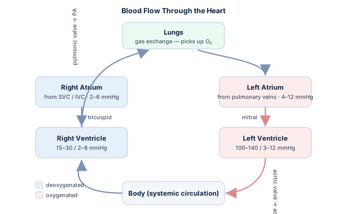
Hemodynamics sits at the center of everything done in a catheterization laboratory, an intensive care unit, and an echo suite. If you are studying the subject from the ground up, our RCIS hemodynamics guide lays out how these measurements connect. This article is educational and is not medical advice — it is meant to build your understanding, not to guide bedside decisions.
Hemodynamics meaning in clinical practice
The textbook hemodynamics meaning is the study of blood flow, but its clinical meaning is broader and more useful: it is the framework clinicians use to answer one question — is this patient's circulation delivering enough oxygenated blood to the tissues? Every hemodynamic measurement is ultimately a proxy for that goal.
Consider how the concept shows up across cardiology. In the cath lab, hemodynamics means the pressures recorded as a catheter advances through the right atrium, right ventricle, pulmonary artery, and wedge position. In the ICU, it means the trend of blood pressure, cardiac output, and filling pressures that guides fluids and vasoactive drugs. In an echo lab, it means the flow velocities and gradients that reveal a stiff valve or a failing ventricle.
- Pressures — the force blood exerts inside each chamber and vessel, measured in millimeters of mercury (mmHg).
- Flow — the volume of blood the heart moves per minute, most importantly cardiac output.
- Resistance — how much the vessels oppose that flow, dominated by the small arterioles.
- Oxygen delivery — the endpoint that all of the above exist to protect.
Because these ideas thread through the entire specialty, a firm grasp of hemodynamics makes the rest of cardiovascular medicine click into place — from cardiac anatomy to the electrical signals covered in our ECG fundamentals guide. The mechanical pump and the electrical wiring are two halves of one organ.
The core principles: pressure, flow, and resistance
Almost all of hemodynamics reduces to one elegant relationship borrowed from electrical physics. Just as Ohm's law states that voltage equals current times resistance, the circulation obeys a hydraulic version of the same law. These are the principles of pressure, flow, and resistance that everything else is built on.
Pressure gradient = Flow × Resistance
Rearranged, flow equals the pressure gradient divided by resistance. In whole-body terms, the mean arterial pressure is roughly the product of cardiac output (flow) and systemic vascular resistance. Change any one variable and the others must move to keep the equation balanced — which is exactly why the body can defend a normal blood pressure even as a disease process quietly shifts the underlying numbers.
| Term | What it is | Typical measure |
|---|---|---|
| Pressure | The driving force behind flow | mmHg |
| Flow | Volume of blood per unit time | L/min (cardiac output) |
| Resistance | Opposition to flow, mostly in arterioles | dynes·sec·cm⁻⁵ |
This single relationship is why the calculated value of systemic vascular resistance depends directly on both blood pressure and cardiac output. It is not measured on its own; it is deduced from the other two. Get comfortable with the triangle and you can reason your way through most hemodynamic problems on the fly.
Pressure: the driving force
Pressure is what gets blood moving. The left ventricle contracts, generates roughly 120 mmHg of systolic pressure, and ejects blood into the aorta; the difference between that high arterial pressure and the near-zero pressure in the veins is the gradient that pushes blood all the way around the loop. Blood, like water, always flows from high pressure to low pressure.
But there is not one pressure — there are many, and each chamber has its own signature. Right atrial pressure sits low (about 2–6 mmHg), the right ventricle generates a modest systolic pressure, the pulmonary artery runs at perhaps 25/10 mmHg, and the left ventricle produces the powerful systemic pressures. Reading these values in sequence as a catheter advances is the heart of an invasive hemodynamic study.
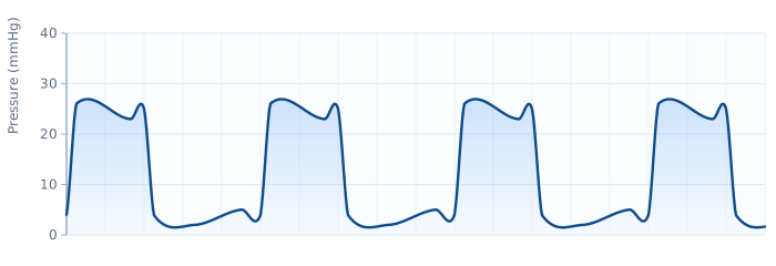
Two summary pressures matter enormously in practice. Mean arterial pressure (MAP) is the average pressure driving blood into the tissues, and it is weighted toward diastole rather than being a simple midpoint of systolic and diastolic. Pulse pressure — the gap between systolic and diastolic — hints at stroke volume and arterial stiffness. Because MAP feeds directly into resistance calculations, and pulse pressure reflects the ejected stroke volume, learning to read pressures is inseparable from learning to read flow.
Flow: cardiac output and its determinants
If pressure is the push, flow is the payoff. The single most important flow measurement in hemodynamics is cardiac output — the volume of blood the heart pumps per minute, normally about 4 to 8 liters. It is the product of two things: how much blood leaves the ventricle per beat (stroke volume) and how many beats occur per minute (heart rate).
Cardiac Output = Stroke Volume × Heart Rate
Stroke volume itself is governed by three classic determinants that every hemodynamics student should be able to recite:
- Preload — the degree of ventricular filling before contraction, reflected in filling pressures. Within limits, more stretch means a more forceful contraction (the Frank-Starling principle).
- Afterload — the resistance the ventricle must overcome to eject, closely tied to vascular resistance.
- Contractility — the intrinsic strength of the heart muscle, independent of loading conditions.
Because output must be fair to compare between a large and a small person, clinicians often index it to body size, yielding the cardiac index. And since stroke volume relative to the total volume in the ventricle gives the familiar ejection fraction, these flow measures interlock. When thermodilution is unreliable, output can be derived from oxygen consumption using the Fick method; our Fick cardiac output calculator shows how that works step by step.
Flow, importantly, is not always smooth. In healthy vessels blood moves in orderly laminar layers, but where a vessel narrows or a valve is stenotic, flow becomes turbulent — chaotic and audible as a murmur or bruit. That transition is the physical basis for much of what we hear with a stethoscope and see on Doppler echocardiography.
Resistance: how vessels shape the circulation
Resistance is the brake on the system. It describes how strongly the blood vessels oppose flow, and it is overwhelmingly controlled by the smallest arteries and arterioles — which is why they are nicknamed the "resistance vessels." Constrict them and pressure rises while flow to that bed falls; dilate them and pressure drops while flow surges.
The physics behind resistance is captured by Poiseuille's law, and its most important lesson is about vessel radius. Resistance is inversely proportional to the fourth power of the radius. That exponent is not a typo: halving a vessel's radius increases its resistance roughly sixteenfold. This is why tiny changes in arteriolar tone produce huge swings in blood pressure, and why vasoconstricting or vasodilating drugs are so powerful.
| Factor | Effect on resistance | Why it matters |
|---|---|---|
| Vessel radius | Resistance ∝ 1/radius⁴ | Dominant factor; small changes have huge effects |
| Vessel length | Longer → more resistance | Relatively fixed in an adult |
| Blood viscosity | Thicker blood → more resistance | Rises with high hematocrit or dehydration |
Two resistances matter clinically: systemic vascular resistance (the afterload for the left ventricle) and pulmonary vascular resistance (the load on the right ventricle). Neither is measured directly — both are calculated from a pressure gradient divided by flow, exactly as the core equation predicts. You can compute them in seconds with our hemodynamic calculator instead of doing the arithmetic by hand.
How hemodynamics is measured in the cath lab
Invasive hemodynamic assessment is where these principles come alive. A thin, fluid-filled or high-fidelity catheter is advanced through the venous or arterial system, and a transducer converts the pressure at its tip into the waveforms you see on the monitor. The classic tool for right-heart hemodynamics is the balloon-tipped Swan-Ganz pulmonary artery catheter, which measures right atrial, right ventricular, pulmonary artery, and wedge pressures, and derives cardiac output by thermodilution.
As the catheter travels from the right atrium onward, each chamber writes its own pressure signature. The wedge (pulmonary capillary) pressure is especially valuable because it approximates left atrial pressure — a downstream window onto the left heart obtained from the right side of the circulation.
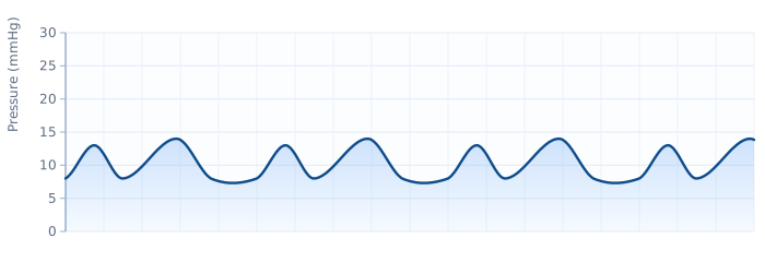
Accuracy hinges on the fundamentals of transducer setup. The transducer must be zeroed to atmospheric pressure and leveled to the phlebostatic axis; an over-damped or under-damped line distorts every reading; and a mistimed thermodilution injection corrupts the cardiac output. Garbage in, garbage out. Before trusting any abnormal value, a good operator confirms the waveform looks clean. To see how invasive study fits alongside intervention, our comparison of PCI versus diagnostic catheterization and the overview of what happens in a cardiac cath lab put the measurements in context.
Hemodynamics in disease: recognizing the patterns
The real power of hemodynamics is diagnostic. Because pressure, flow, and resistance move together in predictable ways, a set of numbers can fingerprint a disease. Shock is the classic teaching example: each type distorts the triangle differently, and the pattern points to the cause.
| Shock type | Cardiac output (flow) | Resistance (SVR) | Filling pressures |
|---|---|---|---|
| Cardiogenic | Low | High | High |
| Hypovolemic | Low | High | Low |
| Distributive (septic) | High / normal | Low | Low |
| Obstructive (tamponade) | Low | High | High & equalized |
Read across any row and the physiology explains itself. A failing pump drops output, so the body constricts vessels (raising resistance) to defend pressure — that is the cold, high-resistance picture of cardiogenic shock hemodynamics. Sepsis does the opposite, dilating vessels until resistance collapses and the patient runs warm despite a low pressure. Obstructive shock, such as cardiac tamponade, produces a distinctive equalization of diastolic pressures as fluid squeezes the heart from outside.
Valve disease writes its own signatures too. A stenotic aortic valve creates a pressure gradient between the left ventricle and the aorta, and the size of that gradient — combined with flow — lets clinicians estimate the aortic valve area. Once you learn to read the pattern rather than any single number, hemodynamics becomes a diagnostic language.
Where hemodynamics meets the electrical heart
Pressure and flow do not happen in a vacuum — they are triggered by the heart's electrical system. Every pressure wave you record is the mechanical echo of an electrical event, and when the electrics go wrong, the hemodynamics follow. This is why hemodynamic and electrophysiologic thinking belong together.
Rhythm is a perfect illustration. In atrial fibrillation, the loss of coordinated atrial contraction removes the "atrial kick" that normally tops off ventricular filling, cutting cardiac output — an effect that worsens at fast rates. A sustained ventricular tachycardia can be so fast and disorganized that the ventricle never fills properly, causing output and pressure to crash. Even a slow rhythm from complete heart block can drop cardiac output enough to require a pacemaker.
The link runs the other way as well: ischemia from a myocardial infarction damages contractility and can wreck hemodynamics within minutes. That is why interpreting the electrical picture is a companion skill, not a separate one. Sharpening your rhythm interpretation and your reading of a STEMI on the ECG pays direct hemodynamic dividends — the wiring and the plumbing fail together.
Studying hemodynamics for the RCIS exam
Hemodynamics is one of the highest-yield domains on the Registered Cardiovascular Invasive Specialist exam and a daily reality for anyone building a career as a cardiovascular technologist. The questions tend to come in three flavors: definitions and concepts, calculations (plug values into an equation), and pattern recognition (match a set of numbers to a diagnosis).
A study sequence that works well is concept, then computation, then application. Start with the definitions and the pressure-flow-resistance triangle until they are automatic. Then drill the core formulas — cardiac output, mean arterial pressure, vascular resistance — until the arithmetic feels effortless; the hemodynamic calculator is a fast way to check your work. Finally, test yourself on scenario questions until you can name a shock type or valve lesion from the numbers alone.
Our RCIS hemodynamics practice questions put these principles into exam-style scenarios, and because waveforms and rhythms are examined side by side, it helps to keep your ECG interpretation skills sharp in parallel. Work the concepts, run the numbers, then apply them under time pressure — that sequence is how the strongest candidates approach the material.
Key takeaways
- Hemodynamics is the study of blood in motion — the pressures, flows, and resistances that keep the tissues perfused.
- One equation rules it all: pressure gradient = flow × resistance, the hydraulic version of Ohm's law.
- Pressure is the driving force, flow is cardiac output (stroke volume × heart rate), and resistance lives mainly in the arterioles.
- Resistance depends on radius to the fourth power, so small changes in vessel caliber cause large changes in pressure and flow.
- Vascular resistance is calculated, not measured — derived from a pressure gradient divided by flow.
- Patterns are diagnostic: shock types and valve lesions each distort pressure, flow, and resistance in recognizable ways.
- Electrical and mechanical function are linked — arrhythmias and infarction disrupt hemodynamics directly.
- Educational only: targets and thresholds continue to evolve; this is study material, not medical advice.
Master hemodynamics
Normal values, waveforms, and cardiac output — the full study guide.
Open the Guide →Frequently asked questions
What is the definition of hemodynamics?
Hemodynamics is the study of the forces that move blood through the circulation. The word comes from the Greek for blood (haima) and force (dynamis). Clinically, it describes the pressures, flows, and vascular resistances that determine how well blood is delivered to the body's organs.
What is the meaning of hemodynamics in simple terms?
In simple terms, hemodynamics means how blood moves and what pushes and resists it. It boils down to three things: the pressure that drives blood forward, the flow (how much blood the heart pumps per minute), and the resistance the vessels put up against that flow. Together they determine whether organs get enough oxygenated blood.
What are the three main principles of hemodynamics?
The three core principles are pressure, flow, and resistance. They are tied together by a hydraulic version of Ohm's law: the pressure gradient equals flow multiplied by resistance. Rearranged, flow equals the pressure gradient divided by resistance. Nearly every hemodynamic calculation is a version of this relationship.
What is the relationship between pressure, flow, and resistance?
Pressure gradient = flow × resistance. Blood flows from high pressure to low pressure, and the vessels' resistance determines how much flow a given pressure produces. If resistance rises and the body wants to keep flow constant, pressure must increase; if resistance falls, pressure drops unless flow increases to compensate.
Why is vessel radius so important to resistance?
Because resistance is inversely proportional to the fourth power of the vessel's radius (Poiseuille's law). Halving the radius increases resistance about sixteenfold. This is why the small arterioles, which can change their caliber dramatically, are the main controllers of blood pressure and regional blood flow.
What is cardiac output and how is it calculated?
Cardiac output is the volume of blood the heart pumps per minute, normally about 4 to 8 liters. It equals stroke volume (the blood ejected per beat) multiplied by heart rate. Its determinants are preload, afterload, and contractility. It can be measured by thermodilution or estimated with the Fick oxygen-consumption method.
How is hemodynamics measured in the cath lab?
Hemodynamics is measured by advancing a pressure-sensing catheter through the heart and vessels. A Swan-Ganz pulmonary artery catheter records right atrial, right ventricular, pulmonary artery, and wedge pressures, and derives cardiac output by thermodilution. Accurate readings depend on properly zeroed, leveled transducers and clean, undamped waveforms.
What does it mean to be hemodynamically stable?
Being hemodynamically stable means the patient's blood pressure and cardiac output are adequate to perfuse vital organs without emergency support. A hemodynamically unstable patient has pressures or flows too low to maintain perfusion, which may require fluids, medications, or mechanical support. Note that this is general education, not medical advice.
Why do arrhythmias affect hemodynamics?
Because the electrical rhythm triggers the mechanical pumping. In atrial fibrillation, the loss of coordinated atrial contraction removes the atrial kick and lowers cardiac output. Very fast rhythms like ventricular tachycardia leave too little time for the ventricle to fill, and very slow rhythms reduce output — all of which disrupt pressure and flow.
Sources & further reading
- Cardiovascular Credentialing International (CCI)
- American College of Cardiology
- American Heart Association
- MedlinePlus (U.S. National Library of Medicine)
External links are provided for reference; always confirm current details with the official source.