Stroke Volume: Normal Range, Formula & What Controls It
Stroke volume is the amount of blood your heart ejects with a single beat — the quiet variable that decides whether every heartbeat delivers a lot of flow or barely enough. This guide covers the normal range, the formula, the three forces that set it, and how it is measured and used at the bedside and in the cath lab.
What is stroke volume?
Stroke volume is the volume of blood the left ventricle ejects into the aorta with each contraction. If cardiac output is the circulation's bottom line, stroke volume is the amount packed into every single beat that builds toward it. A healthy heart does not just beat often enough — it makes each beat count, pushing a meaningful slug of blood forward every time it squeezes.
Picture the left ventricle at the end of filling, brimming with blood. That maximum is the end-diastolic volume. When the ventricle contracts, it does not empty completely; a residual amount, the end-systolic volume, always stays behind. Stroke volume is simply the difference — what actually left the chamber. At rest a typical adult ventricle holds roughly 120 mL at end-diastole and ejects about 70 mL of it, leaving 50 mL behind.
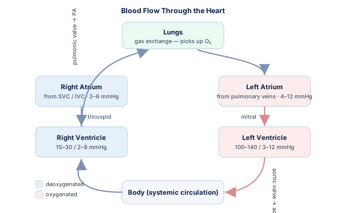
Because stroke volume sits at the center of how the heart performs, it links directly to nearly every other hemodynamic number. If you are studying for a credential exam, it helps to read this alongside our broader hemodynamics study guide, which frames stroke volume within the full picture of pressures, flows, and resistances.
The stroke volume formula
The core definition could not be simpler:
Stroke volume (SV) = end-diastolic volume (EDV) − end-systolic volume (ESV)
In words, stroke volume is what the ventricle held when full minus what it kept after squeezing. Using the typical figures above, 120 mL − 50 mL = 70 mL per beat. That single subtraction is the foundation of the whole topic.
A second relationship ties stroke volume to the number most clinicians actually track. Because cardiac output equals heart rate multiplied by stroke volume, you can always rearrange it:
Stroke volume = cardiac output ÷ heart rate
Stroke volume also anchors two related measures. Divide it by body surface area and you get the stroke volume index, the size-adjusted version. And the fraction of the filled ventricle that gets ejected — stroke volume divided by end-diastolic volume — is the ejection fraction. You can run these and related figures quickly with our hemodynamic calculator.
Normal stroke volume values
The commonly quoted resting adult range for normal stroke volume is 60 to 100 mL per beat, with about 70 mL as the classic average. As with most hemodynamic numbers, the exact figures vary slightly between textbooks and depend on body size, age, sex, and fitness — a large athlete may sit well above 100 mL at rest, while a small adult runs lower.
| Measure | Typical resting range | Note |
|---|---|---|
| Stroke volume | 60–100 mL | Per beat |
| Stroke volume index | 33–47 mL/m² | Stroke volume ÷ body surface area |
| End-diastolic volume | ~120 mL | Left ventricle when full |
| End-systolic volume | ~50 mL | Residual after ejection |
| Ejection fraction | 52–72% (men), 54–74% (women) | Fraction of EDV ejected |
These are educational reference ranges, not treatment thresholds; a real patient is always read in context. Note that stroke volume can be misleadingly reassuring on its own — a dilated, failing ventricle may keep stroke volume near normal by holding a huge end-diastolic volume, even as its ejection fraction falls. That is one reason clinicians rarely look at a single number in isolation; our cardiac index guide explains how flow is size-adjusted for fairer comparison.
Preload, afterload, and contractility
Three physiologic forces set stroke volume, and almost every clinical change in it traces back to one of them. Learning the trio of preload, afterload, and contractility is the single most useful thing you can take from this topic.
Preload
Preload is the degree to which the ventricle is stretched by incoming blood at the end of filling — essentially the end-diastolic volume. By the Frank–Starling mechanism, a more stretched ventricle contracts more forcefully and ejects more, up to a physiologic limit. This is why giving fluid raises stroke volume in a hypovolemic patient and why anything that reduces venous return — bleeding, dehydration, or high intrathoracic pressure — drops it.
Afterload
Afterload is the load the ventricle must work against to open the aortic valve and eject, dominated by arterial pressure and systemic vascular resistance. Raise afterload — severe hypertension, aortic stenosis, intense vasoconstriction — and stroke volume tends to fall because the ventricle meets more resistance. Conversely, vasodilators can improve stroke volume in a failing heart by unloading it.
Contractility
Contractility (inotropy) is the intrinsic strength of the squeeze, independent of how full or how loaded the ventricle is. Sympathetic stimulation, circulating catecholamines, and inotropic drugs such as dobutamine increase it; ischemia, acidosis, beta-blockade, and many cardiomyopathies reduce it. A more contractile ventricle empties more completely, lowering end-systolic volume and raising stroke volume.
Stroke volume variation (SVV)
Stroke volume is not perfectly steady from beat to beat — it rises and falls a little with the breathing cycle, and that swing carries real diagnostic value. Stroke volume variation (SVV) is the percentage difference between the maximum and minimum stroke volume over a respiratory cycle, and it has become one of the most useful bedside tools for predicting whether a patient will respond to fluids.
The physiology is elegant. During positive-pressure ventilation, each mechanical breath transiently squeezes the great veins and reduces venous return, so stroke volume dips slightly, then recovers. In a patient sitting on the steep, preload-dependent part of the Frank–Starling curve — that is, someone who is volume-responsive — that respiratory swing is large. In a well-filled patient on the flat part of the curve, the swing is small because more preload would not help.
| SVV value | Interpretation |
|---|---|
| Under ~10% | Likely NOT fluid-responsive; the ventricle is on the flat part of the curve |
| Around 10–13% | Gray zone — interpret with the full clinical picture |
| Over ~13% | Likely fluid-responsive; a fluid bolus should raise stroke volume |
Because SVV predicts fluid responsiveness more reliably than static pressures like central venous pressure, current critical-care thinking generally favors these dynamic measures for guiding resuscitation, though the ideal threshold and the best monitor are still debated and evolving. It is derived from arterial-waveform pulse-contour analysis, which we return to in the measurement section.
How stroke volume is measured
There is no direct "stroke volume meter." Instead, several methods estimate it from different physical principles, each with its own strengths and blind spots. In practice the choice depends on how invasive the setting allows and how much precision the situation demands.
| Method | How it works | Best used when |
|---|---|---|
| Echocardiography | LVOT cross-sectional area × velocity-time integral (Doppler) | Noninvasive bedside or clinic assessment |
| Thermodilution | Cardiac output ÷ heart rate, via a pulmonary-artery catheter | Routine ICU and cath-lab monitoring |
| Pulse-contour analysis | Beat-to-beat estimate from the arterial pressure waveform | Continuous trending and SVV calculation |
| Cardiac MRI / ventriculography | EDV − ESV from imaged chamber volumes | Precise volumes and research-grade accuracy |
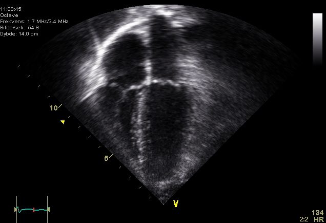
Echocardiography is the workhorse: it measures the diameter of the left ventricular outflow tract to get its cross-sectional area, then uses Doppler to capture the velocity-time integral — how far the column of blood travels per beat — and multiplies the two. It is noninvasive and repeatable, but operator-dependent and sensitive to an accurate LVOT measurement, since that diameter is squared. If sonography interests you, see the role of the cardiovascular ultrasound technologist. In the invasive lab, stroke volume most often falls out of a measured cardiac output divided by heart rate, using a pulmonary-artery catheter.
Low and high stroke volume states
An abnormal stroke volume splits into two clinically opposite pictures. Low stroke volume means each beat delivers too little; high stroke volume reflects either a big, efficient athletic heart or a compensating one.
| Low stroke volume | High stroke volume | |
|---|---|---|
| Common causes | Hypovolemia, heart failure, large myocardial infarction, severe aortic stenosis, cardiac tamponade, high afterload | Athletic conditioning, bradycardia (compensatory), aortic regurgitation, high-output states |
| The mechanism | Poor filling, weak squeeze, or too much resistance to ejection | Large, well-filled or highly contractile ventricle, or slow rate demanding more per beat |
| Typical clues | Narrow pulse pressure, cool extremities, fatigue, low output | Wide pulse pressure, strong bounding pulse, resting bradycardia in athletes |
A trained endurance athlete illustrates the compensatory side neatly: a resting heart rate in the 40s still supports a normal cardiac output because the enlarged, efficient ventricle ejects a large stroke volume with each slow beat. On the failing side, when stroke volume drops far enough that tissues no longer get the oxygen they need, the patient slides toward shock. Conditions that restrict filling, such as cardiac tamponade, or that obstruct ejection, such as a tight aortic valve, both cut stroke volume from different directions.
This is general educational information about physiology, not medical advice; any specific patient's numbers are interpreted by their clinical team alongside pressures, labs, and imaging.
Why stroke volume matters clinically
Stroke volume is not an abstract figure — it is the lever that many bedside decisions actually pull. Because cardiac output is just stroke volume times heart rate, a patient whose stroke volume is collapsing can maintain output only by driving the heart rate up, which is exactly what a compensatory tachycardia represents. Recognizing that a racing pulse may be defending a failing stroke volume changes how you read the whole picture.
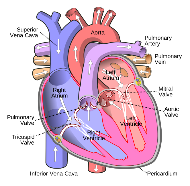
In the cath lab and ICU, stroke volume and its variation steer real actions: whether to give a fluid bolus, whether to start an inotrope to boost contractility, whether to unload a stressed ventricle with a vasodilator, and whether mechanical support such as an intra-aortic balloon pump is needed to prop up each beat. It also feeds derived calculations — cardiac output, cardiac index, and vascular resistance all depend on an accurate stroke volume, so an error here propagates downstream. Understanding the underlying pump helps; our cardiac anatomy guide reviews the chambers and valves involved, and you can drill the concepts in our hemodynamics practice questions.
Key takeaways
- Stroke volume = end-diastolic volume − end-systolic volume, roughly 120 − 50 = 70 mL in a typical resting adult.
- Normal stroke volume is about 60–100 mL per beat; it can also be found as cardiac output divided by heart rate.
- Three forces set it — preload and contractility raise it, while afterload lowers it (remember PAC).
- Stroke volume variation (SVV) predicts fluid responsiveness: over ~13% usually means a fluid bolus will help, but it is only valid in a ventilated, sinus-rhythm patient.
- It is estimated by echocardiography (LVOT area × VTI), thermodilution, pulse-contour analysis, or volumetric imaging — each with distinct strengths.
- Low stroke volume points to poor filling, weak contraction, or high afterload; a high value can reflect an athletic heart or compensation.
- This article is educational and not a substitute for professional medical evaluation.
Calculate stroke volume
Enter cardiac output and heart rate to get stroke volume and its index.
Open the Stroke Volume Calculator →Frequently asked questions
What is a normal stroke volume?
About 60 to 100 mL per beat at rest for an average adult, with roughly 70 mL as the classic textbook value. The exact figure varies with body size, age, sex, and fitness, so it is often indexed to body surface area (33–47 mL/m²) for fairer comparison.
What is the stroke volume formula?
Stroke volume equals end-diastolic volume minus end-systolic volume (SV = EDV − ESV). Using typical values, 120 mL − 50 mL gives about 70 mL. It can also be calculated as cardiac output divided by heart rate.
What are the three determinants of stroke volume?
Preload (how much the ventricle is stretched by incoming blood), afterload (the resistance it must eject against), and contractility (the intrinsic strength of the squeeze). Preload and contractility increase stroke volume, while higher afterload decreases it.
What is stroke volume variation (SVV)?
SVV is the percentage change in stroke volume across a respiratory cycle. In a mechanically ventilated, sinus-rhythm patient, a value above roughly 13% suggests the patient is fluid-responsive and a fluid bolus should raise stroke volume, whereas a value under about 10% suggests it will not.
What is the difference between stroke volume and ejection fraction?
Stroke volume is the actual milliliters of blood ejected per beat, while ejection fraction is the percentage of the filled ventricle that gets ejected — stroke volume divided by end-diastolic volume. A dilated heart can keep a near-normal stroke volume yet have a low ejection fraction.
How is stroke volume measured?
Most commonly by echocardiography, which multiplies the left ventricular outflow tract area by the Doppler velocity-time integral. It can also be derived from thermodilution cardiac output divided by heart rate, from arterial pulse-contour analysis, or from imaged chamber volumes on cardiac MRI or ventriculography.
What causes a low stroke volume?
Hypovolemia and blood loss (low preload), heart failure or a large myocardial infarction (weak contractility), and severe aortic stenosis or intense vasoconstriction (high afterload). Restrictive conditions like cardiac tamponade also lower it by limiting filling.
Why do athletes have a high stroke volume?
Endurance training enlarges and strengthens the left ventricle so it ejects more blood per beat. This large stroke volume lets a well-conditioned athlete maintain a normal cardiac output even with a resting heart rate in the 40s or 50s.
How does heart rate affect stroke volume?
At very high rates, diastole shortens so much that the ventricle has less time to fill, reducing preload and lowering stroke volume — which is why an extremely fast rhythm can drop cardiac output despite the rapid pulse. A slower rate allows fuller filling and a larger stroke volume per beat.
Sources & further reading
- Cardiovascular Credentialing International (CCI)
- American College of Cardiology
- American Heart Association
- MedlinePlus (U.S. National Library of Medicine)
External links are provided for reference; always confirm current details with the official source.