Cardiogenic Shock: Causes, Hemodynamics & Treatment
Cardiogenic shock is what happens when the heart, still full of blood, can no longer push it forward — a lethal traffic jam that leaves the tissues starving while the lungs fill up. Its hemodynamic signature is unmistakable: a low cardiac index sitting on top of a stubbornly high wedge pressure.
- What cardiogenic shock actually is
- Causes of cardiogenic shock
- The pathophysiology: a downward spiral
- The hemodynamic profile: low CI, high PCWP
- Recognizing it at the bedside: cold and wet
- Telling it apart from other shock states
- Treatment: inotropes, IABP, and revascularization
- Monitoring and the role of the cath lab
- Cardiogenic shock on the RCIS exam
- Key takeaways
What cardiogenic shock actually is
Cardiogenic shock is a state of tissue hypoperfusion caused by the heart's own failure to pump — the tank is full, but the pump has quit. Unlike a bleeding patient who has lost volume, the cardiogenic-shock patient usually has plenty of blood; the problem is that a damaged ventricle cannot move it. Oxygen delivery collapses, lactate climbs, the skin turns cold and clammy, urine output falls, and the mind clouds. It is the most lethal of the classic shock states, and even with modern care mortality remains sobering.
Clinically, the working definition combines two things: evidence of low output — a persistently low systolic pressure (often under about 90 mmHg for more than 30 minutes, or the need for support to keep it above that) — plus signs that the tissues are not being perfused despite adequate or elevated filling pressures. That last clause is the key that separates cardiogenic shock from simple hypovolemia: the ventricle is not empty, it is failing. If you want the underlying circulatory physics first, the RCIS hemodynamics guide lays out how pressure, flow, and resistance interlock before you layer a failing pump on top.
This article is educational, written for RCIS candidates, cardiovascular technologists, and clinicians building their foundations. It is not medical advice — real patients are managed by their own care teams using current guidelines and institutional protocols, and the field of shock management continues to evolve.
Causes of cardiogenic shock
The single dominant cause is a large acute myocardial infarction, specifically one that damages enough left-ventricular muscle — classically more than about 40% — that the pump can no longer sustain output. Roughly four out of five cases of cardiogenic shock trace back to acute coronary events, which is why fluency in STEMI ECG interpretation and the wider picture of myocardial infarction is inseparable from managing this shock. The culprit is usually a large anterior infarction from a proximal left anterior descending occlusion, but any territory can do it if enough muscle dies.
Beyond simple pump-muscle loss, MI can cause shock through mechanical complications — and these are the ones that turn a stable patient catastrophic within minutes:
- Acute mitral regurgitation from papillary muscle rupture, flooding the left atrium and lungs.
- Ventricular septal rupture, shunting blood left-to-right and stealing forward output.
- Free-wall rupture, which usually presents as tamponade and sudden death.
Cardiogenic shock is not only an infarct disease, though. Other causes include end-stage dilated cardiomyopathy, fulminant myocarditis, acute valvular failure (severe aortic or mitral disease), arrhythmias — both tachyarrhythmias like ventricular tachycardia and profound bradycardia from complete heart block — as well as right-ventricular infarction, drug toxicity, and stress (takotsubo) cardiomyopathy. A useful way to organize them is by the part of the pump that failed.
| Mechanism | Representative causes |
|---|---|
| Loss of contractile muscle | Large acute MI, myocarditis, end-stage cardiomyopathy |
| Mechanical failure | Papillary muscle rupture (acute MR), VSD, free-wall rupture, acute severe valve disease |
| Rhythm failure | Sustained VT/VF, complete heart block, severe bradycardia |
| Right-heart failure | RV infarction, massive pulmonary embolism (overlaps obstructive) |
The pathophysiology: a downward spiral
What makes cardiogenic shock so deadly is that it feeds on itself. Once the ventricle fails, a vicious cycle takes hold. Falling cardiac output lowers coronary perfusion pressure, which starves the very myocardium that is already struggling, worsening contractility and dropping output further. Meanwhile the body, sensing hypoperfusion, clamps down the arterioles — systemic vascular resistance rises to defend blood pressure, but that added afterload makes the failing ventricle work even harder against a stiffer wall, further reducing forward flow.
At the same time, blood backs up behind the failing left ventricle. Left-atrial and pulmonary venous pressures rise, fluid is forced across the pulmonary capillaries into the alveoli, and the patient develops pulmonary edema — the "wet" half of the classic picture. Hypoxemia from that edema then worsens myocardial oxygen supply, tightening the spiral another turn. Layer on a systemic inflammatory response, which paradoxically can lower SVR in some patients and blur the hemodynamic picture, and you can see why cardiogenic shock resists simple fixes.
The hemodynamic profile: low CI, high PCWP
If you remember one thing about cardiogenic shock, make it this pairing: a low cardiac index sitting on top of a high pulmonary capillary wedge pressure. That combination — poor forward flow despite high left-sided filling pressure — is the fingerprint that separates cardiogenic shock from every other type. A hypovolemic patient also has low output, but their wedge is low; the cardiogenic patient's wedge is high precisely because blood is damming up behind the failing pump.
The defining numbers most consensus definitions use are a cardiac index below roughly 2.2 L/min/m² (often lower, under 1.8, without support) together with a PCWP above about 15–18 mmHg. Central venous pressure typically rises as the right heart congests, and systemic vascular resistance climbs as the body compensates. Understanding why output is scaled to body size is worth a detour through cardiac index, and the raw flow it derives from is covered in cardiac output.
| Parameter | Direction | Typical value | Why |
|---|---|---|---|
| Cardiac index (CI) | ↓ low | < 2.2 L/min/m² | Failing pump can't generate forward flow |
| PCWP (wedge) | ↑ high | > 15–18 mmHg | Blood backs up behind the left ventricle |
| CVP (right preload) | ↑ high / normal | often elevated | Right heart congests as pressures back up |
| SVR (afterload) | ↑ high | > 1200 dynes·s·cm⁻⁵ | Compensatory vasoconstriction |
| Mean arterial pressure | ↓ low | hypotensive | Output falls faster than SVR can compensate |
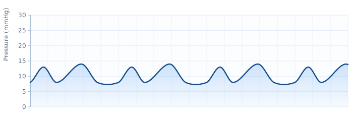
These pressures are read directly from the Swan-Ganz (pulmonary artery) catheter, which floats through the right heart to sample CVP, PA pressures, and the wedge, and measures output by thermodilution. SVR is never measured — it is calculated — and you can watch how the inputs combine in our hemodynamic calculator. How that clamped-down resistance behaves is explored further in systemic vascular resistance.
Recognizing it at the bedside: cold and wet
The classic cardiogenic-shock patient is "cold and wet." Cold, because low output and high SVR leave the skin clamped, mottled, and cool with delayed capillary refill and weak, thready pulses. Wet, because the high wedge pressure has driven fluid into the lungs — crackles, breathlessness, frothy sputum, and hypoxemia. Add hypotension, tachycardia, oliguria, and a dulled sensorium, and the picture assembles quickly.
A widely taught bedside shorthand comes from the Forrester classification, which sorts heart-failure patients into four boxes based on perfusion (cold vs. warm) and congestion (wet vs. dry). Cardiogenic shock lives in the "cold and wet" quadrant — hypoperfused and congested at once — and that box carries the worst prognosis. Not every patient reads the textbook, though: a subset presents "cold and dry" with low output but without florid congestion, and right-ventricular infarction can produce clear lungs with a high CVP and hypotension, a pattern that punishes anyone who reflexively diuretics them.
Telling it apart from other shock states
On the RCIS exam and at the bedside, the fastest way to separate the four shock states is to read cardiac output and SVR together first, then use the filling pressures to pinpoint the type. Cardiogenic and hypovolemic shock both show low output and high SVR — the difference is entirely in the wedge: high in cardiogenic (blood backing up), low in hypovolemic (an empty tank). Distributive (septic) shock runs backward, with low SVR and often high output. The broader four-way framework is laid out in our companion piece on shock hemodynamics.
| Shock type | CVP | PCWP | Cardiac output | SVR |
|---|---|---|---|---|
| Hypovolemic | ↓ | ↓ | ↓ | ↑ |
| Cardiogenic | ↑ | ↑ | ↓ | ↑ |
| Obstructive | ↑ | variable | ↓ | ↑ |
| Distributive | ↓/normal | ↓/normal | ↑ | ↓ |
One overlap deserves care: obstructive shock from cardiac tamponade or massive pulmonary embolism can also show high CVP and low output, mimicking cardiogenic shock. The distinguishing move is the wedge and the pattern of diastolic pressures — in tamponade they equalize, a scenario dissected in cardiac tamponade hemodynamics, while in pulmonary embolism the wedge stays low-normal because the left heart is underfilled behind the clot. In cardiogenic shock, by contrast, the wedge is unambiguously high because the failure sits in the left ventricle itself.
Treatment: inotropes, IABP, and revascularization
Treatment attacks the downward spiral from several directions at once. The interventions fall into three buckets: fix the cause, support the pump, and unload the ventricle.
Revascularization comes first. Because the overwhelming majority of cases stem from acute MI, emergency reopening of the culprit artery — usually primary PCI — is the single intervention with the strongest survival benefit, and current guidance is to revascularize the culprit lesion promptly. Where and how that happens is covered in PCI versus cardiac catheterization and, for the setting itself, what a cardiac cath lab is. Time is muscle; every minute of delay costs myocardium.
Pharmacologic support aims to restore forward flow and, where needed, blood pressure:
- Inotropes such as dobutamine or milrinone increase contractility to squeeze more output from the failing ventricle. They come at a cost — more oxygen demand and arrhythmia risk — so they are used at the lowest effective dose.
- Vasopressors like norepinephrine are added when mean arterial pressure is dangerously low; norepinephrine is often preferred over dopamine, which has been associated with more arrhythmias in shock trials.
- Diuretics and vasodilators help unload the congested ventricle once perfusion allows, though vasodilators must be used cautiously when pressure is marginal.
The interplay of these agents is a recurring cath-lab theme; the cath-lab medications and heart-failure medications overviews are natural companions here.
Mechanical circulatory support steps in when drugs are not enough. The classic device is the intra-aortic balloon pump (IABP), which inflates in diastole to boost coronary perfusion and deflates in systole to lower afterload — elegant physiology, though large trials (notably IABP-SHOCK II) failed to show a mortality benefit in MI-related cardiogenic shock, so its role is now more selective. Newer percutaneous options include axial-flow devices (such as the Impella family), which actively pump blood from the ventricle into the aorta, and veno-arterial ECMO for the most refractory cases. The evidence base for which device helps whom is still maturing, and device selection belongs to the treating team.
Monitoring and the role of the cath lab
Cardiogenic shock is a monitoring-intensive diagnosis. Continuous ECG catches the arrhythmias that both cause and complicate it — sustained supraventricular tachycardia, ventricular arrhythmias, and conduction blocks all show up on the same leads, which is why solid ECG interpretation skills are inseparable from shock care. An arterial line tracks pressure beat to beat, and in complex or mixed presentations a pulmonary artery catheter directly reports the wedge, CVP, PA pressures, and thermodilution output.
Echocardiography is often the fastest bedside tool of all: it shows the failing ventricle, estimates ejection fraction, and rapidly unmasks mechanical complications like acute mitral regurgitation, a ventricular septal defect, or a pericardial effusion causing tamponade. Falling output can be traced to a collapsing stroke volume, and echo helps localize why.
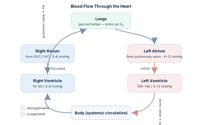
The cath lab is where diagnosis and treatment converge: the same procedure that confirms the culprit coronary lesion also reopens it, and mechanical support devices are placed there under fluoroscopy. Reading the pressure waveforms in real time is a core cath-lab skill; the pressure-tracing work in the hemodynamics guide and hands-on hemodynamics practice questions are the fastest way to build that fluency.
Cardiogenic shock on the RCIS exam
Registry questions on cardiogenic shock are formulaic once you internalize the low-CI/high-PCWP fingerprint. The classic item hands you a set of hemodynamic values and asks you to name the shock type, or gives you a post-MI patient and asks what the wedge, output, and SVR should show. Work it in a fixed order: (1) read cardiac output and SVR — low output with high SVR narrows you to hypovolemic, cardiogenic, or obstructive; (2) read the wedge — a high PCWP with low output points straight at cardiogenic; (3) use the clue words — post-infarct, pulmonary edema, cold-and-wet — to lock it in.
Round out your prep with the flow-and-resistance fundamentals, then drill under timed conditions. The RCIS hemodynamics question bank mixes shock patterns with waveform reading, and pairing it with the ECG guide covers the arrhythmias that so often ride alongside a failing pump. Because cardiogenic shock is fundamentally an infarct disease, time spent on ECG strip practice pays off directly on exam day.
Key takeaways
- Cardiogenic shock is pump failure — the tank is full, but a damaged heart cannot move blood forward, so tissues starve while the lungs flood.
- The dominant cause is acute MI (about 80% of cases), especially large anterior infarcts and mechanical complications like acute MR, VSD, or free-wall rupture.
- The hemodynamic fingerprint is low CI with high PCWP — cardiac index under ~2.2 L/min/m² and wedge above ~15–18 mmHg, with high CVP and high SVR.
- Bedside it is "cold and wet": cold, clamped skin from low output and high SVR; wet lungs from the high wedge.
- High wedge separates it from hypovolemia; both share low output and high SVR, but hypovolemia has a low wedge.
- Treatment attacks the spiral: revascularize first (usually primary PCI), support with inotropes and vasopressors, and unload the ventricle — adding mechanical support (IABP, axial-flow pumps, or ECMO) when drugs fall short.
- Read cardiac output and SVR together first, then use the wedge and clue words to pinpoint the diagnosis.
- This is educational material for exam prep and clinical foundations, not medical advice — real management follows current guidelines and the treating team.
Practise shock hemodynamics
Test shock profiles, waveforms, and support devices.
Practise Hemodynamics →Frequently asked questions
What is cardiogenic shock?
Cardiogenic shock is a life-threatening state in which the heart cannot pump enough blood to meet the body's needs despite adequate or elevated filling pressures. Unlike hypovolemic shock, the problem is not a lack of blood volume but a failing pump. It causes hypotension, cold and clammy skin, pulmonary congestion, poor urine output, and altered mentation, and it remains the most lethal of the classic shock states.
What is the most common cause of cardiogenic shock?
By far the most common cause is a large acute myocardial infarction, which accounts for roughly 80% of cases — typically a big anterior infarct that damages enough left-ventricular muscle to cripple the pump. Mechanical complications of MI (acute mitral regurgitation from papillary muscle rupture, ventricular septal rupture, and free-wall rupture) are also important causes, along with end-stage cardiomyopathy, myocarditis, acute valve failure, and severe arrhythmias.
What is the hemodynamic profile of cardiogenic shock?
The signature is a low cardiac index (below about 2.2 L/min/m²) combined with a high pulmonary capillary wedge pressure (above about 15–18 mmHg). Central venous pressure is usually elevated as the right heart congests, and systemic vascular resistance rises from compensatory vasoconstriction, while mean arterial pressure falls. In short: low forward flow with high left-sided filling pressure.
How do you tell cardiogenic shock from hypovolemic shock?
Both show low cardiac output and high SVR, so the deciding factor is the wedge pressure. Cardiogenic shock has a high PCWP because blood backs up behind the failing ventricle, whereas hypovolemic shock has a low PCWP because the tank is empty. A high wedge with low output means cardiogenic; a low wedge with low output means hypovolemic.
Why is the wedge pressure high in cardiogenic shock?
Because the failure sits in the left ventricle. When the ventricle cannot eject effectively, blood dams up behind it — left-atrial and pulmonary venous pressures rise, and the pulmonary capillary wedge pressure, which estimates left-atrial pressure, climbs. That elevated wedge is what drives fluid into the lungs and produces the pulmonary edema of the classic 'cold and wet' picture.
How is cardiogenic shock treated?
Treatment attacks the cause and the physiology together: emergency revascularization of the culprit coronary artery (usually primary PCI) is the priority and offers the strongest survival benefit; inotropes such as dobutamine or milrinone boost contractility; vasopressors like norepinephrine support blood pressure when needed; and diuretics or vasodilators unload the congested ventricle when perfusion allows. Mechanical circulatory support — an intra-aortic balloon pump, axial-flow devices, or ECMO — is added when drugs are insufficient.
Does an intra-aortic balloon pump improve survival in cardiogenic shock?
The IABP has elegant physiology — it inflates in diastole to augment coronary perfusion and deflates in systole to lower afterload — but large randomized trials, notably IABP-SHOCK II, did not show a mortality benefit in cardiogenic shock from myocardial infarction. As a result its routine use has been downgraded in guidelines, and it is now used more selectively while newer devices are studied. Device choice is individualized by the treating team.
What does 'cold and wet' mean in cardiogenic shock?
'Cold and wet' is the Forrester classification quadrant that cardiogenic shock occupies. 'Cold' refers to hypoperfusion — cool, mottled skin, weak pulses, and low output with high vascular resistance. 'Wet' refers to congestion — pulmonary edema driven by the high wedge pressure, producing crackles, breathlessness, and hypoxemia. Being both hypoperfused and congested carries the worst prognosis of the four quadrants.
What cardiac index defines cardiogenic shock?
Most consensus definitions use a cardiac index below roughly 2.2 L/min/m² with pharmacologic or mechanical support, and often below 1.8 L/min/m² without support, together with a pulmonary capillary wedge pressure above about 15 mmHg. These thresholds capture the essential idea: forward flow is inadequate despite adequate or high filling pressures. Exact cutoffs vary between definitions and continue to be refined.
Sources & further reading
- Cardiovascular Credentialing International (CCI)
- American College of Cardiology
- American Heart Association
- MedlinePlus (U.S. National Library of Medicine)
External links are provided for reference; always confirm current details with the official source.