Supraventricular Tachycardia (SVT): ECG & Treatment
Supraventricular tachycardia is a fast, usually narrow-complex rhythm that originates at or above the AV node — and because it turns a calm monitor into a racing 180-beats-per-minute tracing in an instant, recognizing it and knowing how adenosine ends it are core skills for the cath lab and the RCIS exam.
- What Is Supraventricular Tachycardia?
- Recognizing SVT on the ECG
- Types of SVT: AVNRT, AVRT, and Atrial Tachycardia
- Causes, Triggers, and Risk Factors
- Symptoms and Hemodynamic Impact
- SVT Treatment: Vagal Maneuvers, Adenosine, and Beyond
- Catheter Ablation and Long-Term Management
- SVT in the Cath Lab and on the RCIS Exam
- Key Takeaways
What Is Supraventricular Tachycardia?
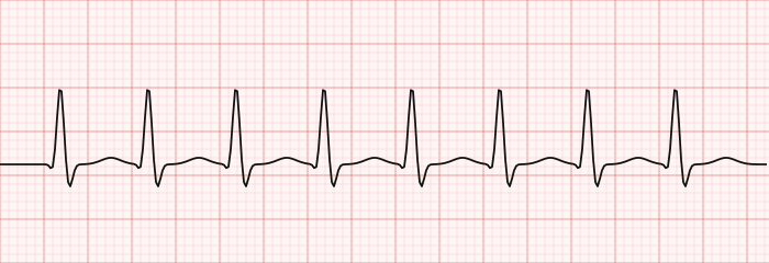
Supraventricular tachycardia (SVT) is an umbrella term for any rapid rhythm that arises from a focus or circuit at or above the level of the AV node — in the atria, the AV junction, or a bypass tract. Because the impulse still travels down the normal, fast His-Purkinje highway in most cases, the ventricles depolarize quickly and in sync, which is why the classic SVT tracing shows a narrow QRS complex at a very fast rate.
In everyday clinical shorthand, "SVT" usually means paroxysmal SVT — the sudden-onset, regular, narrow-complex tachycardias driven by reentry, chiefly AV nodal reentrant tachycardia (AVNRT) and AV reentrant tachycardia (AVRT). Strictly speaking, though, the supraventricular family also includes atrial fibrillation, atrial flutter, and atrial tachycardia. On the RCIS blueprint you are expected to know both the broad definition and the narrow, everyday one.
Rates in SVT typically run 150 to 250 beats per minute. That speed is the whole story clinically: it slashes the time the ventricles have to fill in diastole, so each beat ejects less blood. Understanding how rate and filling drive forward flow is worth reviewing alongside our primers on cardiac output and stroke volume.
This article is educational and written for RCIS exam preparation. It is not medical advice; clinical decisions belong to the treating team following current protocols.
Recognizing SVT on the ECG
Reading SVT ECG findings comes down to a short pattern: narrow, regular, fast complexes that seem to appear from nowhere and often stop just as abruptly. If you want to shore up the fundamentals first, our guides to ECG interpretation and ECG rhythm interpretation walk through the basics that make SVT easier to spot.
Here is what to look for when you suspect SVT:
- Narrow QRS — typically < 120 ms, because conduction to the ventricles is normal.
- Rapid, regular rate — usually 150–250 bpm, with strikingly constant R-R intervals.
- Absent or abnormal P waves — P waves are often hidden inside the QRS or distort its end (a pseudo-R' in V1 or pseudo-S in the inferior leads points to AVNRT).
- Sudden onset and offset — paroxysmal SVT starts and stops abruptly, unlike sinus tachycardia's gentle ramp.
- No warm-up — sinus tachycardia speeds up and slows down gradually; reentrant SVT does not.
A few caveats keep you honest. SVT can occasionally be wide — when it conducts with a bundle-branch block or aberrancy, or down an accessory pathway — and then it mimics ventricular tachycardia. The safe rule for any wide-complex tachycardia is to assume VT unless you are certain otherwise. It is also easy to confuse a fast, regular SVT at exactly 150 bpm with atrial flutter conducting 2:1; slowing AV conduction with a vagal maneuver or adenosine often unmasks the flutter waves.
Types of SVT: AVNRT, AVRT, and Atrial Tachycardia
Most exam questions and most real cases boil down to a handful of mechanisms. The two reentrant heavyweights — AVNRT AVRT — account for the large majority of paroxysmal SVT, and they are worth distinguishing because they behave and are ablated differently.
AV nodal reentrant tachycardia (AVNRT) is the single most common type. It uses two functionally distinct pathways within the AV node — a slow pathway and a fast pathway — that form a tiny reentrant loop. A well-timed premature beat blocks in one pathway and circles down the other, and the loop sustains itself. Because the atria and ventricles are activated almost simultaneously, the P wave is usually buried in the QRS. AVNRT skews toward younger and middle-aged patients, more often women, and frequently occurs in structurally normal hearts.
AV reentrant tachycardia (AVRT) uses an accessory pathway — an extra strand of muscle connecting atrium to ventricle outside the AV node. In orthodromic AVRT (the common form), the impulse goes down the AV node and back up the accessory pathway, producing a narrow complex. In the rarer antidromic form, it goes the other way and the QRS is wide. When the accessory pathway also shows up on the resting ECG as a short PR and a delta wave, the patient has Wolff-Parkinson-White (WPW) pattern — clinically important because certain drugs are dangerous in WPW with atrial fibrillation.
Atrial tachycardia arises from an ectopic atrial focus firing rapidly, independent of the AV node. It often shows a P-wave morphology different from sinus and does not necessarily terminate with adenosine, which helps separate it from the AV-node-dependent reentrant tachycardias.
| Feature | AVNRT | AVRT | Atrial tachycardia |
|---|---|---|---|
| Circuit location | Within AV node | Accessory pathway + AV node | Ectopic atrial focus |
| Typical P wave | Buried in QRS (pseudo-R' in V1) | Retrograde, after QRS | Abnormal, before QRS |
| Adenosine terminates? | Usually yes | Usually yes | Often no (transient block) |
| Associated with WPW? | No | Yes (delta wave possible) | No |
| Relative frequency | Most common | Second most common | Less common |
Causes, Triggers, and Risk Factors
Unlike ventricular tachycardia, paroxysmal SVT often strikes in structurally normal hearts. The underlying substrate — dual AV nodal pathways or an accessory pathway — is usually present from birth, so many patients describe episodes going back years. What varies is what tips a quiet substrate into a run of tachycardia.
Common triggers and contributors include:
- Premature beats — an atrial or ventricular extrasystole timed just right initiates reentry.
- Stimulants — caffeine, nicotine, some decongestants, and illicit stimulants.
- Adrenergic surges — stress, exertion, pain, or fever raising catecholamines.
- Alcohol and dehydration.
- Thyroid disease — hyperthyroidism lowers the threshold for atrial arrhythmia.
- Electrolyte disturbances and, in some people, hormonal shifts.
Procedural triggers matter to the cath-lab tech too. Catheter contact with the atrial wall, guidewire manipulation, and contrast injection can provoke atrial ectopy that launches a short SVT run. Patients arriving with an acute coronary syndrome or in the cardiac cath lab for other reasons may reveal a previously silent pathway under the stress of the procedure. Knowing the patient's baseline rhythm and keeping crash equipment ready is standard lab discipline.
Symptoms and Hemodynamic Impact
How a patient feels in SVT tracks with the rate, its duration, and the health of the underlying heart. Most people describe a sudden pounding or fluttering in the chest, and the abrupt on-off quality is a strong clue that the rhythm is reentrant rather than sinus. Along with palpitations, patients commonly report lightheadedness, shortness of breath, chest discomfort, anxiety, and sometimes a strong urge to urinate afterward (from atrial natriuretic peptide release during the episode).
The hemodynamic problem is mostly one of rate. At 180 to 220 beats per minute, diastole is so short that ventricular filling suffers and stroke volume drops, so even a normal heart can see its blood pressure sag. Loss of a well-timed atrial contraction — the atrial "kick" — compounds it. In a heart with limited reserve, a low ejection fraction, or significant coronary disease, a fast SVT can precipitate angina, heart failure, or, rarely, cardiogenic shock. Reviewing how pressure and flow interact in our hemodynamics guide makes it clearer why a young patient tolerates 200 bpm while an older one with ischemia does not.
Clinically you are triaging one question above all: is the patient stable or unstable? Signs of instability include hypotension, altered mental status, ischemic chest pain, and acute heart failure. That single determination steers the entire treatment algorithm below.
SVT Treatment: Vagal Maneuvers, Adenosine, and Beyond
The first branch point in acute management is the familiar one: is the patient stable or unstable? Current resuscitation and arrhythmia guidance builds the algorithm around that answer.
Unstable SVT with a pulse — hypotension, ischemic chest pain, altered mentation, or acute heart failure — calls for prompt synchronized cardioversion, with sedation if time allows.
Stable, regular narrow-complex SVT gives you room to work stepwise. Start with vagal maneuvers, which increase parasympathetic tone and can break AV-node-dependent reentry. The Valsalva maneuver is first-line, and the modified Valsalva — blowing into a syringe while supine, then having the legs raised as you lie the patient flat — converts substantially more patients than the standard technique in trial data. Carotid sinus massage is an alternative in appropriate patients after listening for bruits.
When vagal maneuvers fail, SVT treatment adenosine is the pharmacologic workhorse. Adenosine briefly blocks conduction through the AV node — the very tissue the reentrant circuit depends on — and so terminates AVNRT and orthodromic AVRT in most cases. It is given as a rapid IV push followed by an immediate saline flush, because its half-life is only seconds. The typical adult dose is 6 mg, then 12 mg if needed. Patients should be warned about the brief but intense feeling of chest pressure, flushing, or impending doom that accompanies the drug's fleeting asystolic pause.
| Scenario | First-line action |
|---|---|
| Unstable SVT, has pulse | Synchronized cardioversion |
| Stable regular narrow SVT | Vagal maneuvers (modified Valsalva) |
| Vagal maneuvers fail | IV adenosine 6 mg, then 12 mg |
| Adenosine fails / recurs | IV calcium-channel or beta-blocker |
| Irregular wide SVT (WPW + AF) | Procainamide / cardioversion — avoid AV nodal blockers |
If adenosine does not hold, rate-controlling agents such as intravenous diltiazem, verapamil, or a beta-blocker are reasonable next steps for stable patients. A crucial safety point: in a patient with pre-excited atrial fibrillation (WPW with an irregular, wide, fast rhythm), AV-nodal-blocking drugs — adenosine, calcium-channel blockers, beta-blockers, digoxin — can be dangerous, because they can push conduction down the accessory pathway and accelerate the ventricular rate. Those patients are managed with procainamide or cardioversion instead. The agents used in the lab to manage these rhythms are collected in our cath-lab medications reference.
Catheter Ablation and Long-Term Management
Acute termination is only half the story. For patients with recurrent, symptomatic SVT, the definitive treatment is catheter ablation, which targets the arrhythmia's substrate directly. In AVNRT, the operator modifies the slow pathway; in AVRT, the accessory pathway is ablated. Success rates are high and complication rates low, which is why current guidance offers ablation as a first-line option for many patients with recurrent AVNRT or AVRT rather than a last resort after failed drugs.
For patients who prefer to avoid a procedure, or between episodes, chronic drug therapy is an option. Beta-blockers and non-dihydropyridine calcium-channel blockers (diltiazem, verapamil) reduce episode frequency by slowing AV nodal conduction. Some patients with infrequent, well-tolerated episodes are taught a "pill-in-the-pocket" approach or simply how to perform vagal maneuvers at home. As always, the choice is individualized and belongs to the treating team.
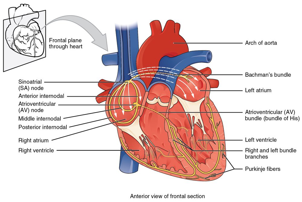
Patients with WPW deserve special mention, because a small subset are at risk of sudden death if atrial fibrillation conducts rapidly down the accessory pathway. Risk stratification — sometimes with an electrophysiology study — and ablation of the pathway are the mainstays. For device and mapping background, our overviews of the pacemaker and the cardiac conduction system put the electrophysiology in context.
SVT in the Cath Lab and on the RCIS Exam
For the registered cardiovascular invasive specialist, SVT is not just a textbook rhythm — it can flash onto the monitor mid-procedure. Catheter contact with the atrial wall, wire manipulation, and adrenergic stress can all launch a run of narrow-complex tachycardia. Most self-terminate or respond to vagal maneuvers and adenosine, but the tech should recognize the rhythm, know that a rapid narrow-complex tracing is usually supraventricular, and be ready to assist.
That reality shapes lab readiness: continuous rhythm monitoring, IV access for a rapid adenosine push with immediate flush, and a defibrillator on standby for the rare unstable case or the wide, irregular WPW-plus-atrial-fibrillation pattern that must never receive AV nodal blockers. If you support hemodynamic assessment, being fluent with the Swan-Ganz catheter helps you read how a sustained tachycardia is affecting filling pressures and output.
On the exam itself, expect to distinguish SVT from sinus tachycardia and atrial flutter, to separate ventricular tachycardia from SVT with aberrancy, to place AVNRT versus AVRT, and to pick the correct first action based on stability. To drill these, work through our targeted RCIS ECG practice questions and the ECG strip identification set, which include narrow- and wide-complex tachycardias alongside heart block and other rhythms.
Key Takeaways
- Supraventricular tachycardia arises at or above the AV node and is usually a fast, regular, narrow-complex rhythm; treat any wide-complex tachycardia as VT until proven otherwise.
- On the SVT ECG, look for a narrow QRS, a very regular rate of 150–250 bpm, absent or buried P waves, and sudden onset and offset.
- AVNRT AVRT are the two dominant reentrant types: AVNRT reenters inside the AV node, while AVRT uses an accessory pathway outside it (delta wave suggests WPW).
- SVT often occurs in structurally normal hearts and is triggered by premature beats, stimulants, stress, or adrenergic surges.
- Management hinges on stability: cardiovert the unstable patient; for stable SVT use vagal maneuvers first, then SVT treatment adenosine, then rate-controlling drugs.
- Avoid AV nodal blockers (including adenosine) in pre-excited atrial fibrillation (WPW); use procainamide or cardioversion instead.
- Catheter ablation is a high-success, often first-line definitive treatment for recurrent AVNRT and AVRT.
- This is educational content for RCIS preparation, not medical advice.
Frequently asked questions
What is supraventricular tachycardia in simple terms?
It is a fast heart rhythm that starts in the upper chambers of the heart or the AV junction — above the ventricles. Because the electrical signal usually reaches the ventricles through the normal pathway, the ECG shows narrow QRS complexes at a very fast, regular rate, commonly 150 to 250 beats per minute. Episodes often start and stop suddenly.
What is the difference between AVNRT and AVRT?
AVNRT (AV nodal reentrant tachycardia) uses two pathways inside the AV node itself to form a small reentry loop, and it is the most common type of paroxysmal SVT. AVRT (AV reentrant tachycardia) needs an accessory pathway — an extra electrical connection outside the AV node linking atrium to ventricle. AVRT is the mechanism behind many Wolff-Parkinson-White cases and may show a delta wave on the resting ECG.
How does adenosine work for SVT?
Adenosine briefly blocks conduction through the AV node, which is the tissue the reentrant circuit depends on. By interrupting that circuit for a few seconds it terminates AVNRT and orthodromic AVRT in most patients. It is given as a rapid IV push with an immediate saline flush because it is broken down within seconds, and patients often feel a short but intense wave of chest pressure or flushing.
How do you tell SVT from sinus tachycardia?
Sinus tachycardia warms up and slows down gradually, has normal P waves before each QRS, and usually stays below about 150 beats per minute in adults. Paroxysmal SVT starts and stops abruptly, is often faster, tends to be strikingly regular, and frequently has P waves that are hidden in or distort the QRS. Vagal maneuvers or adenosine slow sinus tachycardia only transiently but can terminate reentrant SVT.
Is supraventricular tachycardia dangerous?
For most people SVT is fast but not immediately life-threatening, and many episodes resolve with vagal maneuvers or medication. It becomes dangerous when the rate is very high in a patient with limited cardiac reserve, causing low blood pressure, chest pain, or heart failure, or in Wolff-Parkinson-White syndrome where atrial fibrillation can conduct rapidly down an accessory pathway. Unstable patients are treated urgently with synchronized cardioversion.
What are vagal maneuvers and do they work?
Vagal maneuvers increase parasympathetic tone to slow AV node conduction and can break reentrant SVT. The Valsalva maneuver is first-line, and a modified version — blowing into a syringe while lying back, then having the legs raised — converts more patients than the standard technique. Carotid sinus massage is another option in suitable patients after checking for bruits.
Can supraventricular tachycardia be cured?
In many cases, yes. Catheter ablation targets the specific circuit — modifying the slow pathway in AVNRT or eliminating the accessory pathway in AVRT — with high success and low complication rates. It is now offered as a first-line option for many patients with recurrent symptomatic SVT rather than only after medications fail.
Why can adenosine be dangerous in Wolff-Parkinson-White syndrome?
In a patient with WPW who is in atrial fibrillation, the rhythm is irregular and wide because impulses can travel down the accessory pathway. AV-nodal-blocking drugs such as adenosine, calcium-channel blockers, beta-blockers, and digoxin can push more conduction down that pathway and dangerously speed up the ventricular rate. These patients are treated instead with procainamide or cardioversion.
Can SVT happen during a cardiac catheterization?
Yes. Catheter contact with the atrial wall, wire manipulation, contrast injection, and the adrenergic stress of the procedure can trigger a run of supraventricular tachycardia. Most episodes self-terminate or respond to vagal maneuvers and adenosine, but continuous rhythm monitoring and a defibrillator on standby are standard cath-lab precautions.
Sources & further reading
- Cardiovascular Credentialing International (CCI)
- American College of Cardiology
- American Heart Association
- MedlinePlus (U.S. National Library of Medicine)
External links are provided for reference; always confirm current details with the official source.