QT Interval & QTc: Normal Values, Correction & Prolonged QT
The QT interval is the stretch of the ECG where the ventricles power up and then reset — and when that reset drags on, the heart becomes electrically unstable. Read the corrected value, the QTc, correctly and you catch one of the few ECG findings that can genuinely precede a cardiac arrest. This guide is educational and not a substitute for individualized medical advice.
- What Is the QT Interval?
- How to Measure the QT Interval Correctly
- Normal QT and QTc Values
- How the QTc Is Corrected for Heart Rate
- What Prolongs the QT Interval?
- Why a Long QT Matters: Torsades de Pointes
- Managing a Prolonged QT and Torsades
- The QT Interval in the Cath Lab and on the RCIS Exam
- Key Takeaways
What Is the QT Interval?
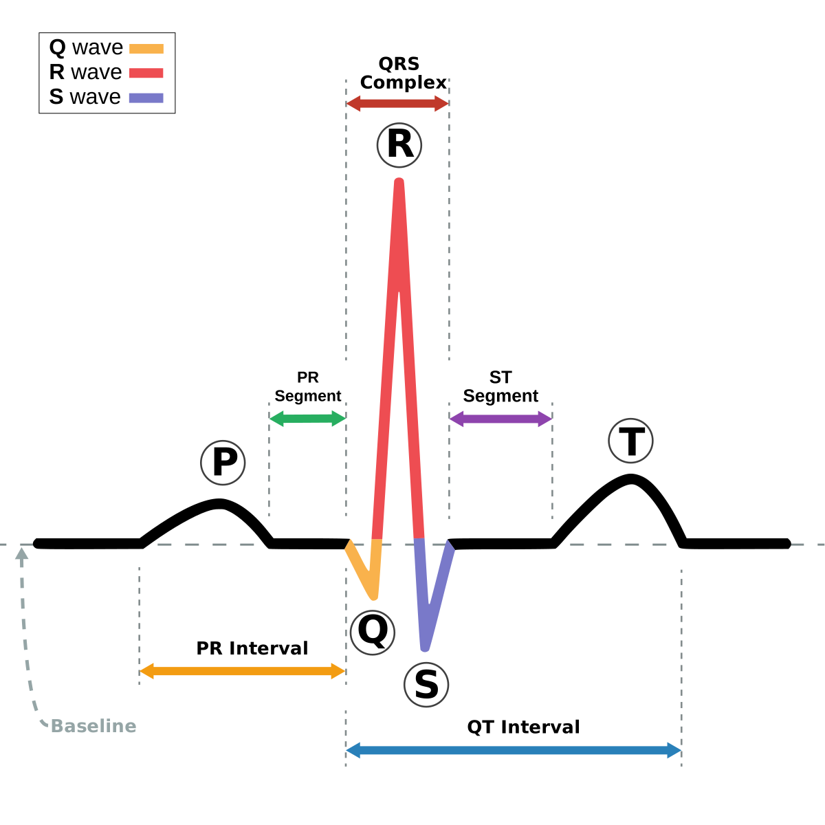
The QT interval is the time from the very start of the QRS complex to the end of the T wave. In plain terms, it captures the full cycle of ventricular electrical activity: depolarization (the QRS, when the ventricles fire and contract) plus repolarization (the ST segment and T wave, when the cells recharge and relax). Nothing else on the surface ECG tells you so directly how long the ventricular muscle stays electrically "busy" before it is ready to beat again.
That window matters because repolarization is the vulnerable phase of the cardiac cycle. If it takes too long, cells recover at uneven rates, and that patchwork of ready and not-ready tissue is exactly the substrate that lets a dangerous rhythm ignite. So while the QT looks like a simple measurement — one point to another — it is really a readout of ventricular repolarization stability.
One complication built into the measurement: the QT is heart-rate dependent. It naturally shortens as the heart speeds up and lengthens when it slows. A raw QT of 400 ms means very different things at 50 beats per minute versus 120. That is why clinicians almost never quote the raw QT alone; they correct it for rate and report the QTc. Understanding the intervals of the ECG — the PR, the QRS, and the QT — is foundational to reading any strip, and it is worth pairing this article with a broader review of ECG interpretation fundamentals.
How to Measure the QT Interval Correctly
Measurement sounds trivial until you actually try it on a real tracing. The start point is easy: the earliest deflection of the QRS from the baseline, in whichever lead shows it first. The end point — where the T wave finishes — is where measurement goes wrong most often.
The accepted technique is the tangent method. Draw a line along the steepest downslope of the T wave and extend it to the isoelectric baseline; the point where that tangent crosses the baseline is the end of the T wave. This gives a reproducible endpoint even when the T wave tails off gradually.
- Pick the right leads. Leads II and V5 are traditional favorites because they usually show a clear T-wave terminus. Guidance suggests measuring in several leads and using the longest reliable value.
- Exclude the U wave. A U wave is a small, later deflection following the T wave. Including it artificially lengthens your QT. If T and U merge, measure to the nadir of the trough between them where you can.
- Average a few beats. The QT varies slightly beat to beat, so measuring three to five consecutive complexes and averaging reduces error.
- Beware automated readings. Machine-calculated QTc is a useful screen but can be fooled by artifact, prominent U waves, or wide QRS complexes. When the number looks alarming, confirm it by hand.
Wide QRS complexes — from bundle branch block, ventricular pacing, or a wide-complex tachycardia such as ventricular tachycardia — inflate the QT simply because depolarization itself is prolonged. In those settings, specialized correction formulas exist, and the raw number should be interpreted with caution.
Normal QT and QTc Values
Because the QT is reported after rate correction, the numbers you memorize are QTc values, expressed in milliseconds. Thresholds differ modestly by sex, since women have slightly longer baseline QTc than men.
| QTc range | Men | Women | Interpretation |
|---|---|---|---|
| Short | < 350 ms | Abnormally short (consider short QT syndrome) | |
| Normal | < 440 ms | < 460 ms | Reassuring |
| Borderline | 440–460 ms | 460–480 ms | Watch and re-check |
| Prolonged | > 480 ms | Abnormal — investigate cause | |
| Markedly prolonged | > 500 ms | High torsades risk | |
Two numbers are worth carrying in your head above all others. A QTc over 500 ms is the widely cited danger line where the risk of torsades de pointes climbs sharply. And an increase of more than 60 ms above a patient's own baseline is a red flag even if the absolute value has not yet crossed 500 — the change matters as much as the ceiling.
A commonly overlooked point: an unusually short QTc is also abnormal. Congenital short QT syndrome and states such as hypercalcemia or digoxin effect shorten repolarization and carry their own arrhythmia risk. Most day-to-day concern, though, centers on the long end of the spectrum.
How the QTc Is Corrected for Heart Rate
Rate correction converts the measured QT into what it "would be" at a heart rate of 60 bpm, so that values are comparable across patients and across time. Several formulas exist, and knowing their quirks keeps you from being misled.
- Bazett: QTc = QT / √RR. Historically the default and still the most widely used, but it over-corrects at fast heart rates (falsely long) and under-corrects at slow rates (falsely short). Its weakness is greatest exactly where you most need accuracy.
- Fridericia: QTc = QT / RR1/3. More reliable across a wide range of rates and increasingly favored, especially in drug-safety work and tachycardia.
- Framingham: a linear correction (QTc = QT + 0.154 × (1 − RR)) that also performs well at rate extremes.
- Hodges: another rate-based linear formula sometimes used in research settings.
In every formula, RR is the R-R interval in seconds — the time between two consecutive R waves, equal to 60 divided by the heart rate. Guidance increasingly recommends Fridericia or Framingham over Bazett when the heart rate is far from 60, precisely because Bazett distorts those readings. For most patients near a normal rate, the formulas converge and the choice barely matters.
This rate dependence is why the QT is inseparable from an understanding of the cardiac conduction system: the same rate that the sinus node and AV node set is the rate that reshapes the repolarization window you are trying to measure.
What Prolongs the QT Interval?
Causes of QT prolongation fall into two broad camps: acquired (something you can often reverse) and congenital (a genetic ion-channel disorder). In hospital practice the acquired causes dominate, and they frequently stack on top of one another.
Medications are the single biggest acquired driver. Many drug classes block the potassium channels responsible for repolarization:
- Antiarrhythmics such as sotalol, amiodarone, dofetilide, and procainamide
- Macrolide and fluoroquinolone antibiotics (azithromycin, levofloxacin)
- Antifungals such as fluconazole
- Antipsychotics and some antidepressants (haloperidol, methadone, citalopram)
- Antiemetics such as ondansetron and droperidol
Electrolyte disturbances are the other heavyweight, and they are common in acutely ill patients:
- Hypokalemia (low potassium)
- Hypomagnesemia (low magnesium)
- Hypocalcemia (low calcium)
Additional contributors include bradycardia, myocardial ischemia, hypothermia, hypothyroidism, and increased intracranial pressure. The danger, clinically, is additive risk: a patient on a QT-prolonging antibiotic who then becomes hypokalemic from diuresis can tip from borderline to dangerous quickly.
| Category | Representative causes |
|---|---|
| Drugs | Sotalol, amiodarone, dofetilide, macrolides, fluoroquinolones, haloperidol, methadone, ondansetron |
| Electrolytes | Low K⁺, low Mg²⁺, low Ca²⁺ |
| Cardiac | Bradycardia, ischemia, heart block |
| Systemic | Hypothermia, hypothyroidism, raised intracranial pressure |
| Congenital | Long QT syndrome (LQTS types 1–3 and others) |
Congenital long QT syndrome (LQTS) is an inherited channelopathy that predisposes to syncope and sudden death, often provoked by specific triggers — exercise or swimming in LQT1, sudden loud noise or emotional startle in LQT2, and sleep or rest in LQT3. It should be considered in anyone with a long QTc, unexplained fainting, or a family history of sudden death.
Why a Long QT Matters: Torsades de Pointes
A prolonged QT is not dangerous in itself — it is dangerous because of what it can trigger. Delayed repolarization allows a phenomenon called early afterdepolarization, a small extra electrical bump during the recovery phase. If that bump arrives during the vulnerable window and lands on the tail of the preceding T wave (the classic "R-on-T" event), it can launch torsades de pointes.
Torsades de pointes — French for "twisting of the points" — is a distinctive form of polymorphic ventricular tachycardia. On the monitor the QRS complexes appear to spiral around the baseline, their amplitude waxing and waning as if the axis were rotating. It is one of the most recognizable and most feared rhythms in cardiology.
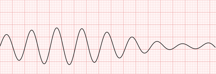
Torsades often self-terminates and produces only palpitations, dizziness, or a brief loss of consciousness. But it can also degenerate into ventricular fibrillation and sudden cardiac arrest. That unpredictability is precisely why a QTc over 500 ms demands attention rather than a wait-and-see approach. Recognizing this rhythm is a core skill in any thorough ECG rhythm interpretation workflow, and it belongs beside your knowledge of other malignant ventricular rhythms.
Managing a Prolonged QT and Torsades
Management splits cleanly into two questions: how do you handle a prolonged QT before anything happens, and what do you do when torsades actually strikes? This section is educational; real cases should follow institutional protocols and clinician judgment.
Preventing an event. The core moves are unglamorous but effective:
- Identify and stop, or substitute, any QT-prolonging drug.
- Replete electrolytes aggressively — keep potassium and magnesium in the higher normal range.
- Continue telemetry monitoring and re-check the QTc after each intervention.
- Avoid stacking additional QT-prolonging agents; check every new medication against a drug database.
Treating active torsades. When the rhythm appears:
- Intravenous magnesium sulfate is first-line, even when the serum magnesium is normal — it stabilizes the myocardium and suppresses the afterdepolarizations that perpetuate torsades.
- Correct potassium and other electrolytes urgently.
- For a pause-dependent, bradycardia-driven torsades, increasing the heart rate shortens the QT — achieved with an infusion of isoproterenol or with temporary transvenous pacing. Faster rates mechanically compress the repolarization window.
- If the patient is unstable or the rhythm degenerates into ventricular fibrillation, defibrillate immediately and run standard resuscitation.
For patients with congenital LQTS, longer-term management extends to beta-blockers, activity or trigger modification, and — in higher-risk cases — an implantable defibrillator, decisions made in specialist cardiology and often electrophysiology clinics.
The QT Interval in the Cath Lab and on the RCIS Exam
For cardiovascular technologists and RCIS candidates, the QT interval is not an abstract curiosity — it shows up in real procedural settings and reliably on the exam. Several catheterization and electrophysiology scenarios intersect directly with QT physiology.
- Contrast and electrolyte shifts. Diuresis, fluid loss, and osmotic effects around a procedure can drop potassium and magnesium, nudging a borderline QT into the danger zone.
- Procedural medications. Sedatives, antiemetics such as ondansetron, and certain antibiotics given periprocedurally can prolong the QT. Cross-referencing the medications commonly used in the cath lab against QT risk is part of safe monitoring.
- Ablation and EP studies. During a cardiac ablation, catheter manipulation and induced rhythms make continuous QT and repolarization awareness essential.
- Recognition on telemetry. The technologist watching the monitor is often the first to see a lengthening QTc or the opening beats of polymorphic VT.
On the exam, expect questions on normal QTc thresholds, the Bazett formula, the “longest reliable lead” measurement principle, magnesium as first-line torsades therapy, and the classic list of QT-prolonging drugs and electrolytes. Because this material overlaps heavily with rhythm recognition, it pays to drill it alongside the rest of your ECG preparation. Our RCIS ECG practice questions and the dedicated ECG strip identification set reinforce exactly these patterns, and the broader RCIS ECG study guide ties the intervals together with rhythm interpretation.
Key Takeaways
- The QT interval spans ventricular depolarization and repolarization, measured from the start of the QRS to the end of the T wave.
- Always report the rate-corrected QTc; use the tangent method, the longest reliable lead, and exclude the U wave.
- Normal QTc is roughly < 440 ms in men and < 460 ms in women; a QTc over 500 ms or a rise of more than 60 ms from baseline signals high risk.
- Bazett is the classic correction but distorts at rate extremes; Fridericia or Framingham are more reliable when the heart rate is far from 60.
- The big drivers are QT-prolonging drugs and low potassium, magnesium, and calcium — remember “low and slow” — plus congenital long QT syndrome.
- A long QT threatens life through torsades de pointes, a polymorphic VT that can progress to cardiac arrest.
- Intravenous magnesium is first-line for torsades; correct electrolytes, stop culprit drugs, and speed the rate for pause-dependent cases.
- This article is for education and exam preparation and is not medical advice.
Calculate QTc
Enter the QT interval and heart rate for the Bazett and Fridericia corrected QT.
Open the QTc Calculator →Frequently asked questions
What is a normal QT interval?
Reported as the rate-corrected QTc, a normal value is under 440 ms in men and under 460 ms in women. Values of 440–480 ms are borderline depending on sex, and a QTc above 480 ms is prolonged. Because the raw QT changes with heart rate, the corrected QTc is the number that actually matters.
What is the difference between QT and QTc?
QT is the interval you measure directly on the ECG. QTc is that value mathematically corrected for heart rate so it can be compared fairly across different rates and patients. Since the raw QT shortens as the heart speeds up, only the QTc lets you judge whether repolarization is truly prolonged.
What is a dangerous QTc?
A QTc above 500 ms markedly raises the risk of torsades de pointes and is the widely cited danger threshold. Just as important, an increase of more than 60 ms above a patient's own baseline is a warning sign even if the absolute value has not yet reached 500 ms.
How is the QT interval corrected for heart rate?
With formulas that use the R-R interval in seconds. Bazett (QT divided by the square root of RR) is the most common but over-corrects at fast rates and under-corrects at slow ones. Fridericia (QT divided by the cube root of RR) and Framingham are more accurate when the heart rate is far from 60 bpm.
What causes a prolonged QT interval?
The leading causes are QT-prolonging medications — certain antiarrhythmics, macrolide and fluoroquinolone antibiotics, some antipsychotics and antidepressants, methadone, and ondansetron — plus low potassium, magnesium, or calcium. Bradycardia, ischemia, hypothermia, hypothyroidism, and congenital long QT syndrome also contribute, and these risks stack on one another.
Why is a long QT dangerous?
A prolonged QT reflects delayed and uneven repolarization, which allows early afterdepolarizations to trigger torsades de pointes — a polymorphic ventricular tachycardia. Torsades can cause fainting and may deteriorate into ventricular fibrillation and sudden cardiac arrest, which is why a very long QTc is treated as urgent.
How do you treat torsades de pointes?
Intravenous magnesium sulfate is first-line, even when serum magnesium is normal. Correct potassium and other electrolytes, stop any offending drug, and for pause-dependent torsades speed the heart rate with isoproterenol or temporary pacing to shorten the QT. An unstable patient or one who deteriorates into ventricular fibrillation needs immediate defibrillation.
How do you measure the QT interval accurately?
Measure from the start of the QRS to the end of the T wave using the tangent method — drawing a line down the steepest part of the T wave's downslope to the baseline. Use a lead with a clear T-wave endpoint such as II or V5, exclude any U wave, and average several beats. Confirm alarming automated readings by hand.
What is a short QT interval and does it matter?
Yes. A QTc under about 350 ms is abnormally short and can indicate congenital short QT syndrome, hypercalcemia, or digoxin effect. Short QT syndrome carries its own risk of arrhythmia and sudden death, so an unusually short value deserves evaluation, though prolonged QT is the far more common clinical concern.
Sources & further reading
- Cardiovascular Credentialing International (CCI)
- American College of Cardiology
- American Heart Association
- MedlinePlus (U.S. National Library of Medicine)
External links are provided for reference; always confirm current details with the official source.