Premature Ventricular Contractions (PVCs): ECG, Causes & When to Worry
Premature ventricular contractions are the extra, early beats almost every heart throws now and then — usually harmless, occasionally a warning, and a near-guaranteed sight on RCIS strips and cath-lab monitors, so knowing how to read them and when to worry is essential.
What Are Premature Ventricular Contractions?
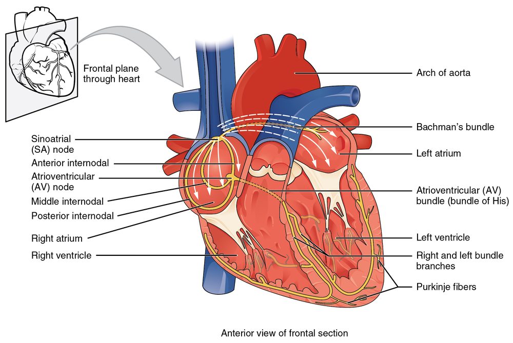
A premature ventricular contraction (PVC) is a heartbeat that fires early from an ectopic focus below the AV node — somewhere in the ventricular muscle or the Purkinje network — rather than from the sinus node. Because the impulse skips the fast His-Purkinje highway and spreads cell-to-cell through myocardium, the ventricles depolarize slowly and out of sequence. That is why a PVC shows up on the tracing as a wide, bizarre-looking QRS complex that arrives sooner than the next expected sinus beat.
PVCs go by many names: ventricular ectopy, ventricular premature beats, ventricular extrasystoles. Whatever the label, they are extraordinarily common. Prolonged monitoring picks them up in the large majority of healthy adults, and their frequency rises with age, caffeine, stress, and structural heart disease. Most people feel nothing at all; some feel a distinct 'skipped beat,' a thud, or a flip-flop in the chest — which is actually the forceful sinus beat that follows the pause, not the PVC itself.
The clinical weight of a PVC depends less on the single beat and more on its company: how often it comes, whether it clusters into patterns, and what kind of heart it lands in. A rare PVC in a structurally normal heart is a non-event. Frequent PVCs in a scarred or weakened ventricle are a different conversation. Understanding how each beat contributes to forward flow is worth reviewing alongside our primers on stroke volume and cardiac output.
This article is educational and written for RCIS exam preparation. It is not medical advice; clinical decisions belong to the treating team following current guidelines.
Recognizing PVCs on the ECG
Spotting a PVC ECG pattern comes down to a short, reliable checklist. The premature beat interrupts the underlying rhythm with a QRS that looks nothing like the patient's normal complexes. If you want to firm up the fundamentals first, our guides to ECG interpretation and ECG rhythm interpretation lay the groundwork that makes ectopy easy to catch.
Here is what defines a PVC on the strip:
- Premature — the beat arrives earlier than the next expected sinus QRS.
- Wide QRS — typically ≥ 120 ms, with a bizarre, un-sinus-like shape.
- No preceding P wave — the impulse starts in the ventricle, so there is no related atrial activity ahead of it.
- Discordant T wave — the T wave usually points opposite to the main QRS deflection.
- Compensatory pause — a full pause often follows, because the sinus node keeps its own timing and the next sinus beat lands 'on schedule.'
The compensatory pause is a useful tell. In a classic PVC the interval spanning the beat before and the beat after the PVC equals two normal R-R intervals — the sinus node was never reset. That distinguishes many PVCs from premature atrial beats, which usually reset the sinus node and produce a shorter, non-compensatory pause. Interpolated PVCs are the exception: they squeeze in between two sinus beats without any pause at all.
Morphology carries information. A PVC with a left-bundle-branch-block pattern and inferior axis often originates in the right ventricular outflow tract — a common, generally benign site. Recognizing these clues is exactly the kind of skill the exam rewards, and you can drill it on our ECG strip identification set.
PVC Patterns: Bigeminy, Trigeminy, Couplets and Runs
PVCs rarely stay solo, and the pattern they fall into has its own vocabulary — vocabulary the RCIS blueprint expects you to know cold. The patterns describe how the ectopic beats interleave with the normal ones.
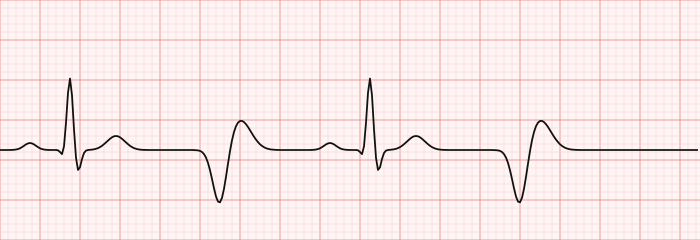
The core terms are worth memorizing:
| Term | Pattern |
|---|---|
| Bigeminy | Every other beat is a PVC (sinus – PVC – sinus – PVC) |
| Trigeminy | Every third beat is a PVC |
| Quadrigeminy | Every fourth beat is a PVC |
| Couplet | Two PVCs in a row |
| Triplet / salvo | Three PVCs in a row (technically a run of nonsustained VT) |
| Unifocal | All PVCs share the same shape (one focus) |
| Multifocal | PVCs of differing shapes (multiple foci) |
Bigeminy is the pattern students remember best because it is so visually distinctive: a metronome-like alternation of a normal beat and an early wide beat. It can look alarming, but ventricular bigeminy is often benign and asymptomatic. One practical caveat — during bigeminy the PVC may not generate a palpable pulse, so the measured heart rate can appear roughly half the true electrical rate. Always correlate the monitor with the patient.
The line where patterns become worrying is well defined. Three or more consecutive PVCs at a rate above 100 bpm is, by definition, a run of ventricular tachycardia. So a triplet is not just extra ectopy — it is the shortest possible VT. Multifocal PVCs and couplets tend to signal a more irritable myocardium than uniform, isolated beats.
PVC Causes and When to Worry
This is the question patients and exam candidates ask most: PVC causes, and when to worry. The reassuring headline is that in a structurally normal heart, occasional PVCs are considered benign. The nuance is in the triggers, the frequency, and the setting.
Common, reversible triggers include:
- Stimulants — caffeine, nicotine, alcohol, and some decongestants or energy drinks.
- Catecholamine surges — stress, anxiety, exercise, and sleep deprivation.
- Electrolyte disturbances — low potassium and low magnesium in particular.
- Hypoxia and stimulant medications — including some asthma inhalers and thyroid excess.
Structural and ischemic causes carry more weight. PVCs are more frequent and more meaningful in the presence of coronary artery disease, prior myocardial infarction, cardiomyopathy, valvular disease, and reduced ejection fraction. During an acute coronary event, new frequent ventricular ectopy can be an early flag of an irritable, ischemic ventricle.
So when should PVCs prompt concern? Current guidance points to several red flags rather than a single cutoff:
- Symptoms — syncope, near-syncope, or PVCs that coincide with chest pain or breathlessness.
- Known heart disease — PVCs on a background of low EF, prior MI, or cardiomyopathy.
- High burden — a large fraction of total beats being ectopic on 24-hour monitoring; a very high burden can, over time, drive a reversible drop in pumping function known as PVC-induced cardiomyopathy.
- Complex patterns — multifocal PVCs, frequent couplets, or runs (nonsustained VT), especially with exertion.
- PVC landing on the T wave — the 'R-on-T' phenomenon, which in a vulnerable heart can trigger dangerous ventricular arrhythmia.
Where exactly the burden threshold for harm sits is still an area of evolving evidence, and estimates vary; the practical point is that a persistently high burden warrants evaluation of ventricular function rather than reassurance alone. For a patient in the cardiac cath lab with an evolving STEMI, new ectopy is watched closely because the ischemic ventricle is primed for trouble.
Symptoms and Hemodynamic Impact
Most PVCs are silent. When symptoms do occur, patients most often describe palpitations: a skipped beat, a pause, a hard thump, or a fluttering sensation. Counterintuitively, the sensation usually comes not from the PVC itself but from the compensatory pause and the extra-forceful sinus beat that follows it, which fills a fuller ventricle and ejects harder.
The hemodynamic effect of a single PVC is small and transient. Because the PVC fires early, the ventricle has had less filling time, so that particular beat ejects a reduced stroke volume and may not produce a palpable pulse. In frequent bigeminy this can meaningfully lower the effective pulse rate and, occasionally, blood pressure. Reviewing how filling and rate drive forward flow in our hemodynamics guide makes it clear why an occasional dropped beat is harmless but a sustained high burden is not.
Over the long term, a very high PVC burden can uncouple the timing of the ventricles and, in some patients, produce a cardiomyopathy with a falling ejection fraction. The encouraging part is that this form is frequently reversible: suppress or ablate the PVCs and pumping function often recovers. That reversibility is precisely why burden quantification on ambulatory monitoring matters.
Diagnostic Workup
The workup for PVCs scales to the clinical picture. A healthy, asymptomatic person with rare PVCs needs little beyond reassurance and attention to triggers. A symptomatic patient, or anyone with red-flag features, gets a more structured evaluation aimed at two questions: how many, and is the heart normal?
- 12-lead ECG — confirms the ectopy, characterizes morphology (hinting at the site of origin), and screens for ischemia, prior infarct, or a long QT.
- Ambulatory monitoring — 24- to 48-hour Holter or longer patch monitoring quantifies the PVC burden as a percentage of total beats and catches patterns like couplets and runs.
- Echocardiogram — assesses ventricular function and structure; a normal study is reassuring, while reduced function reframes the ectopy. See our overview of the echocardiogram for what it measures.
- Exercise testing — PVCs that worsen with exertion or recovery deserve closer attention than those that vanish with faster sinus rates.
- Bloodwork — electrolytes and thyroid function to catch reversible drivers.
When ischemia is suspected as the driver, further evaluation may lead toward angiography and a discussion of PCI versus diagnostic catheterization. Burden and morphology together also guide whether a patient might benefit from ablation, discussed below.
Treatment and Management
Management follows a simple principle: treat the patient and the underlying heart, not the ECG artifact. Asymptomatic PVCs in a structurally normal heart generally need no treatment beyond reassurance and removing obvious triggers such as excess caffeine, alcohol, and stimulants, plus correcting any electrolyte abnormality.
When PVCs are symptomatic or the burden is high, a stepped approach applies:
- Lifestyle and reversible causes — reduce stimulants, address sleep and stress, correct low potassium or magnesium, and treat thyroid excess.
- Beta-blockers — the usual first-line drug for symptom control; calcium-channel blockers are an alternative in selected patients.
- Antiarrhythmic drugs — reserved for more troublesome cases and used cautiously, weighing side effects against benefit.
- Catheter ablation — for a high burden, drug-refractory symptoms, or PVC-induced cardiomyopathy, targeted catheter ablation of the ectopic focus is effective and often curative, particularly for outflow-tract PVCs.
The medications used in the lab to stabilize irritable ventricles are collected in our cath-lab medications reference. Where PVCs coexist with reduced ejection fraction or heart failure, treatment overlaps with standard heart-failure medications, because improving the failing substrate can itself calm the ectopy. For patients whose PVC-driven cardiomyopathy is identified early, the outlook after suppression or ablation is generally favorable — though the field continues to refine which patients benefit most.
PVCs in the Cath Lab and on the RCIS Exam
For the registered cardiovascular invasive specialist, PVCs are not a textbook curiosity — they scroll across the monitor during real procedures. Advancing a catheter into the right or left ventricle mechanically irritates the endocardium and reliably produces PVCs or short runs that stop the instant the catheter is pulled back. Contrast injection into a coronary artery, guidewire contact, and reperfusion after opening an occluded vessel are all classic moments for a burst of ventricular ectopy.
That reality shapes lab discipline: continuous rhythm monitoring, corrected electrolytes before the case, a defibrillator within reach, and an operator who watches the trace and communicates. A few catheter-induced PVCs are expected and benign; a run that organizes into sustained VT is not, and the team must be ready to act. Familiarity with the cardiac conduction system and with mechanical support such as the intra-aortic balloon pump rounds out the picture for higher-risk patients.
On the exam itself, expect to identify PVCs by their premature wide morphology, name the patterns (bigeminy, trigeminy, couplets, runs), distinguish unifocal from multifocal ectopy, and recognize when three-in-a-row becomes nonsustained VT. Comparing PVCs with other rhythms — atrial fibrillation, supraventricular tachycardia, and heart block — is fair game. To drill all of this, work through our targeted RCIS ECG practice questions, and reinforce the underlying anatomy with the ECG guide and cardiac anatomy guide.
Key Takeaways
- A PVC is an early beat from a ventricular focus that produces a wide, bizarre QRS with no preceding P wave and, usually, a compensatory pause.
- On the PVC ECG, look for a premature wide complex with an opposite-direction T wave; the compensatory pause helps separate it from premature atrial beats.
- Patterns matter: bigeminy is every other beat, trigeminy every third, couplets are two in a row, and three or more consecutive PVCs is a run of nonsustained VT.
- For PVC causes and when to worry: isolated PVCs in a normal heart are benign; red flags are syncope, known structural disease, high burden, complex patterns, and the R-on-T phenomenon.
- A very high PVC burden can cause a reversible cardiomyopathy, which is why ambulatory monitoring quantifies burden and echocardiography checks function.
- Management treats the patient and the heart: remove triggers, correct electrolytes, use beta-blockers for symptoms, and consider ablation for refractory or cardiomyopathy-causing ectopy.
- In the cath lab, catheter contact and reperfusion routinely provoke PVCs that resolve on withdrawal — monitoring and defibrillator readiness are standard.
- This is educational content for RCIS preparation, not medical advice.
Practise ectopic-beat recognition
Spot PVCs, bigeminy, and runs on real strips.
Practise ECG Strips →Frequently asked questions
Are premature ventricular contractions dangerous?
In a structurally normal heart, occasional PVCs are considered benign and often need no treatment. They become more concerning when they are frequent, occur in someone with known heart disease or a low ejection fraction, cause symptoms like fainting, or appear in complex patterns such as couplets, runs, or multifocal beats. The company a PVC keeps matters more than the single beat.
What does a PVC look like on an ECG?
A PVC appears as an early, wide, and oddly shaped QRS complex with no P wave in front of it, and the T wave usually points opposite to the main deflection of the QRS. It is typically followed by a full compensatory pause, because the sinus node keeps its own timing and the next sinus beat lands on schedule.
What is ventricular bigeminy?
Bigeminy is a repeating pattern in which every other beat is a PVC — one sinus beat, then one PVC, over and over. It can look dramatic on the monitor but is often benign and asymptomatic. One practical note: the PVC in bigeminy may not produce a palpable pulse, so the felt heart rate can appear about half the true electrical rate.
When should I worry about PVCs?
Seek evaluation if PVCs come with fainting or near-fainting, chest pain, or breathlessness; if you have known heart disease or reduced heart function; if monitoring shows a high burden; or if they occur in complex patterns like frequent couplets, runs, or multifocal beats. Isolated PVCs in a healthy, asymptomatic person usually do not require concern.
What causes premature ventricular contractions?
Common triggers include caffeine, nicotine, alcohol, stress, poor sleep, and stimulant medications, along with low potassium or magnesium and thyroid excess. More significant causes include coronary artery disease, prior heart attack, cardiomyopathy, and reduced ejection fraction, where PVCs tend to be both more frequent and more meaningful.
How many PVCs per day is too many?
There is no single agreed cutoff, and the evidence is still evolving. Rather than a fixed number, clinicians look at the PVC burden as a percentage of total beats on 24-hour monitoring. A persistently high burden warrants an assessment of heart function, because over time it can drive a reversible drop in pumping ability known as PVC-induced cardiomyopathy.
Can PVCs cause heart failure?
A very high burden of PVCs can, in some patients, lead to a cardiomyopathy with a falling ejection fraction. The good news is that this form is frequently reversible — suppressing the PVCs with medication or eliminating the focus with catheter ablation often allows heart function to recover. This is a key reason why high-burden ectopy is worked up rather than ignored.
What is the difference between a PVC and a run of ventricular tachycardia?
A single PVC is one early wide beat. When three or more PVCs occur consecutively at a rate above 100 beats per minute, it meets the definition of ventricular tachycardia — so a triplet is essentially the shortest possible VT run. Two in a row is called a couplet, which sits between an isolated PVC and a true run.
Do PVCs need treatment?
Often not. Asymptomatic PVCs in a normal heart are usually managed with reassurance and by removing triggers such as excess caffeine and correcting electrolytes. When PVCs cause bothersome symptoms or reach a high burden, beta-blockers are the usual first-line therapy, and catheter ablation is highly effective for refractory cases or PVC-induced cardiomyopathy.
Sources & further reading
- Cardiovascular Credentialing International (CCI)
- American College of Cardiology
- American Heart Association
- MedlinePlus (U.S. National Library of Medicine)
External links are provided for reference; always confirm current details with the official source.