Cardiac Catheterization Complications
Cardiac catheterization is one of the safest invasive procedures in modern medicine, but it is not risk-free. Knowing the complications cold — how to spot them, why they happen, and what to do next — is core knowledge for anyone working in the cath lab or preparing for the RCIS exam.
- Cardiac cath complications: the big picture
- Access site complications
- Retroperitoneal bleed: the hidden emergency
- Dissection and vessel injury
- Cardiac complications: arrhythmias, MI, and tamponade
- Contrast nephropathy and kidney risk
- Stroke, allergic reaction, and radiation
- Who is high risk — and how complications are prevented
- Key takeaways
Cardiac cath complications: the big picture
Cardiac cath complications are the adverse events that can occur during or after a diagnostic or interventional catheterization. The reassuring headline first: serious complications are uncommon. For a routine diagnostic coronary angiogram in a stable patient, the risk of a major event — death, stroke, myocardial infarction, or a vascular injury needing repair — is well under one percent. Interventional procedures such as PCI carry somewhat higher risk because they involve balloons, stents, and longer catheter dwell times, but even then the odds strongly favor the patient.
Still, "uncommon" is not "never," and the cath lab is stocked and staffed precisely because when something goes wrong it can go wrong quickly. The most useful mental model is to group complications by where they happen: at the access site where the sheath enters the artery, in the vasculature the catheters travel through, in the heart itself, in the kidneys that clear the contrast, and as systemic events like stroke or allergic reaction. Every complication in this article fits into one of those buckets.
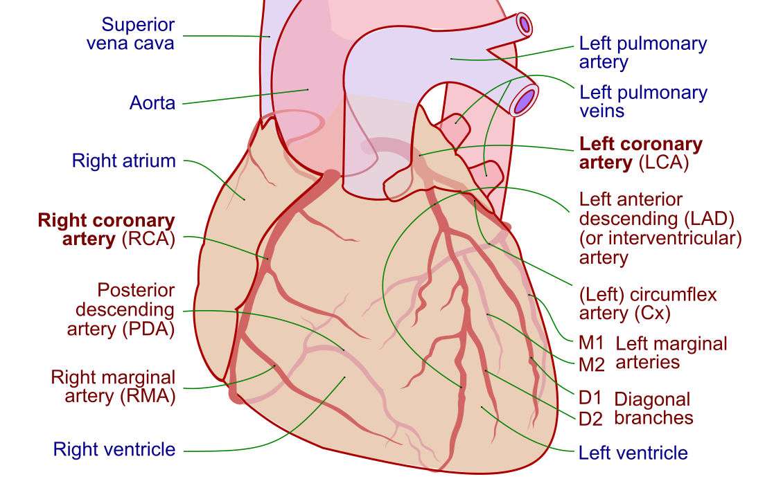
Risk is not evenly distributed. A young, stable patient having an elective diagnostic study sits at the low end; an elderly patient in cardiogenic shock undergoing emergency PCI sits at the high end. Recognizing which patients are high-risk before the case even begins is half the battle. This article is educational, written for students and clinicians preparing for exams such as the RCIS — it is not medical advice or a procedural protocol. Always follow your institution's current policies and the operator's judgment.
Access site complications
The access site is where the sheath punctures the artery, and it is the single most common source of complications after cardiac catheterization. That makes sense: it is the one place where a large hole is deliberately made in a pressurized artery and then has to be closed again. The choice between the radial artery in the wrist and the femoral artery in the groin shapes the entire risk profile.
The field has shifted decisively toward radial-first access over the past two decades, and access-site bleeding is the main reason why. The wrist is superficial and easy to compress, the artery is small, and there is no deep space for blood to hide. Femoral access, by contrast, sits over the pelvis where a leak can bleed inward, unseen, into the retroperitoneum. Current interventional guidance favors the radial approach for most patients specifically because it reduces major bleeding and vascular complications.
The main femoral access-site complications to know are:
- Hematoma — a localized collection of blood under the skin. Common, usually minor, managed with pressure, but a large or expanding one is a warning sign.
- Pseudoaneurysm — blood tracks out of the artery into a contained pocket that still communicates with the lumen, producing a pulsatile mass and often a to-and-fro bruit. Diagnosed by ultrasound and treated with compression or thrombin injection.
- Arteriovenous (AV) fistula — an abnormal channel between the punctured artery and an adjacent vein, classically causing a continuous murmur.
- Arterial occlusion or thrombosis — more relevant to the smaller radial artery, where the vessel can clot off; usually asymptomatic thanks to the dual blood supply of the hand, but a recognized event.
- Infection — uncommon with sterile technique but possible, especially with prolonged sheath dwell times.
Retroperitoneal bleed: the hidden emergency
Of all the access-site complications, the retroperitoneal bleed deserves its own section because it is the one that hides. When a femoral puncture is placed too high — above the inguinal ligament, over the pelvic brim — blood can leak backward into the retroperitoneal space instead of pooling visibly under the skin. There is no obvious groin hematoma to see. The patient can lose a large volume of blood into that space before anyone notices, which is exactly what makes it dangerous.
The presentation is often deceptively quiet at first, then abrupt. Classic clues include:
- Unexplained hypotension and tachycardia after the procedure, out of proportion to anything visible.
- Back, flank, or lower abdominal pain, sometimes with groin or thigh discomfort.
- A falling hemoglobin and hematocrit without an obvious external source of bleeding.
- Diaphoresis, restlessness, or a vasovagal-like picture that does not fully add up.
Because the blood is hidden, the diagnosis rests on suspicion plus imaging — usually a CT scan — and the management is resuscitation: stop or reverse anticoagulation, give fluids and blood as needed, and in severe cases pursue endovascular or surgical control. A high femoral stick is the leading risk factor, which is why meticulous puncture technique (and increasingly, ultrasound guidance and the radial approach) is the best prevention.
Recognizing this picture draws directly on an understanding of the hemodynamics of shock — a patient who is bleeding into the retroperitoneum is heading toward hypovolemic shock, and the falling blood pressure and rising heart rate are the body's early compensation. Sharpening those instincts is exactly what working through hemodynamics practice questions is for.
Dissection and vessel injury
Dissection is a tear in the inner lining of a blood vessel that lets blood burrow into the vessel wall, creating a false channel. In the cath lab this can happen anywhere a catheter, wire, or balloon touches an artery — at the access site, along the aorta, at the tip of a guiding catheter engaging a coronary ostium, or within a coronary artery during PCI. A dissection can be trivial and self-sealing, or it can be catastrophic if it propagates or occludes flow.
Coronary artery dissection is the one that grabs everyone's attention. During PCI, a balloon or stent can split the vessel wall; a guiding catheter can dissect the ostium of the left main or right coronary artery. If the false lumen expands and compresses the true lumen, blood flow stops and the patient can infarct on the table. This is why operators watch the contrast column obsessively — a dissection often announces itself as a lingering, hazy stain of dye or a sudden change in flow. Most procedure-related coronary dissections are managed by stenting across the tear to tack the flap back against the wall.
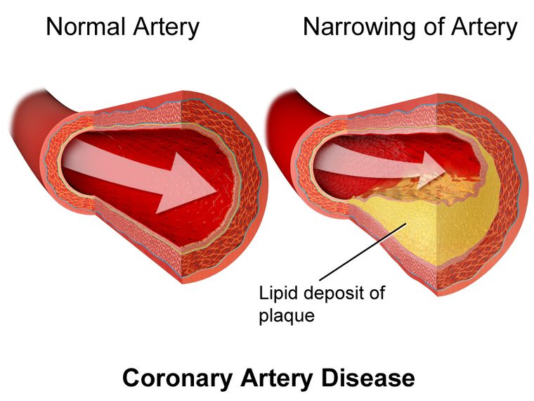
Aortic dissection is a rarer but feared event, usually from catheter manipulation at the aortic root or coronary ostia, and it can be immediately life-threatening. Coronary perforation — a full-thickness hole rather than a wall split — is a related and dangerous complication of interventional work, particularly with stiff wires, oversized balloons, or atherectomy. A perforation can bleed into the pericardium and cause tamponade within minutes.
Because these events unfold as changes in coronary flow and on the ECG, confident ECG interpretation and quick recognition of an evolving ST-elevation MI pattern are part of catching them early. A working knowledge of the coronary artery anatomy being instrumented — which vessel supplies what territory — turns an abstract complication into a concrete, anticipatable risk.
Cardiac complications: arrhythmias, MI, and tamponade
Putting catheters into a beating heart inevitably provokes it. Most of the cardiac complications are transient and benign, but a few are genuine emergencies, and the cath lab team has to tell them apart in real time.
Arrhythmias are the most frequent cardiac events. Catheter contact with the myocardium triggers ectopy; contrast injection can slow the heart; and reperfusion during PCI can set off dangerous rhythms. The spectrum runs from harmless premature beats to life-threatening chaos:
- Bradycardia and transient heart block, often from contrast injection or catheter contact, especially in the right coronary artery.
- Atrial fibrillation and other supraventricular rhythms provoked by catheter irritation of the atria.
- Ventricular tachycardia and ventricular fibrillation — the feared rhythms, more likely during reperfusion of an occluded artery or with ischemia. These demand immediate defibrillation.
This is why continuous rhythm monitoring runs throughout every case, and why fluency in rhythm interpretation is non-negotiable at the bedside. Recognizing the difference between a run of ventricular tachycardia that needs the defibrillator and a self-limited burst of ectopy is a skill built by repetition — the kind you get from ECG strip practice.
Periprocedural myocardial infarction can result from dissection, thrombus, distal embolization of plaque debris, or acute closure of a treated vessel. Cardiac tamponade — blood filling the pericardial sac and squeezing the heart — is the most time-critical cardiac complication, usually following a coronary perforation or, rarely, chamber puncture during a right heart study. The hemodynamic signature is unmistakable once you know it: rising heart rate, falling blood pressure, pulsus paradoxus, and equalization of diastolic pressures across the chambers. Recognizing it on the tracing is a classic teaching point covered in depth in our review of the hemodynamics of cardiac tamponade, and it is treated emergently with pericardiocentesis.
| Cardiac complication | Typical trigger | Immediate response |
|---|---|---|
| Bradycardia / heart block | Contrast injection, RCA catheter contact | Cough, atropine, temporary pacing |
| VT / VF | Reperfusion, ischemia, catheter contact | Defibrillation, ACLS |
| Periprocedural MI | Dissection, thrombus, vessel closure | Restore flow, stent, adjunct drugs |
| Tamponade | Perforation, chamber puncture | Pericardiocentesis, reverse anticoagulation |
Contrast nephropathy and kidney risk
Contrast nephropathy — increasingly called contrast-associated acute kidney injury — is the deterioration of kidney function in the days after a contrast-based procedure. It is classically defined as a rise in serum creatinine (for example, a 25% relative or 0.5 mg/dL absolute increase) within 48 to 72 hours of contrast exposure, with no other explanation. The creatinine typically peaks at three to five days and, in most patients, recovers.
An honest caveat the field has been wrestling with: the true independent contribution of contrast to kidney injury is smaller than older literature suggested. Much of that early data could not separate the effect of the dye from the underlying illness, dehydration, and other nephrotoxins in sick patients. Modern controlled data suggest the risk attributable to contrast alone is modest in many patients — a reason for calibrated caution, not for withholding a needed angiogram.
Risk is concentrated where several factors stack up:
- Pre-existing chronic kidney disease — by far the biggest driver, especially at low eGFR.
- Diabetes, particularly with coexisting kidney disease.
- Dehydration or reduced effective circulating volume.
- Heart failure and low cardiac output states.
- Large contrast volumes and closely spaced repeat studies.
The most evidence-backed protection is simple and cheap: intravenous isotonic hydration before and after the procedure in at-risk patients, combined with using the smallest effective contrast volume. Older adjuncts such as N-acetylcysteine and sodium bicarbonate infusions have not held up in large trials and are no longer broadly recommended. Because poor pump function feeds kidney risk, this topic ties back to a working grasp of cardiac output — a struggling circulation and a contrast load are a combination worth respecting. Our full article on iodinated contrast media covers the dye chemistry and allergy side of the story in depth.
Stroke, allergic reaction, and radiation
A handful of complications are systemic — they affect the whole patient rather than a single vessel or chamber. They are individually uncommon but important, and they round out the differential when a patient does poorly after a case.
Stroke is among the most feared cath complications because it can be devastating and permanent. It usually results from embolization — a fragment of atherosclerotic plaque, a thrombus, or air dislodged as catheters and wires cross the aortic arch near the vessels that feed the brain. Most periprocedural strokes are ischemic and embolic; a small number are hemorrhagic, related to the anticoagulation used during the case. Any new focal neurologic deficit during or after catheterization — weakness, facial droop, slurred speech, visual loss — must be treated as a stroke until proven otherwise and triggers immediate neurologic evaluation.
Allergic and anaphylactoid reactions to iodinated contrast range from mild hives to, rarely, full anaphylaxis with airway compromise and circulatory collapse. Confusingly, most are not true IgE-mediated allergies, but the emergency response is the same regardless. Patients with a documented prior contrast reaction are premedicated with a corticosteroid and antihistamine, though premedication reduces rather than eliminates risk — which is why every injection happens where anaphylaxis can be treated.
Radiation injury is the quiet, cumulative complication. Fluoroscopy delivers ionizing radiation to the patient's skin and to the staff standing beside the table for years. Long, complex interventions can, rarely, deliver enough skin dose to cause a radiation burn. Protection rests on the classic principles — minimizing fluoroscopy time, using collimation and low frame rates, maximizing distance, and disciplined shielding for staff. Air embolism and cholesterol (atheroembolic) embolization, where showers of cholesterol crystals lodge in small vessels of the skin, kidneys, or gut, round out the systemic hazards.
Who is high risk — and how complications are prevented
Complications are far more predictable than random. Certain patients walk into the lab already carrying elevated risk, and identifying them is the foundation of prevention. High-risk features include advanced age, chronic kidney disease, diabetes, peripheral arterial disease, severe left ventricular dysfunction, prior stroke, uncontrolled hypertension, and the acutely unstable — patients in cardiogenic shock or with an evolving myocardial infarction. Emergency and complex interventional procedures carry more risk than elective diagnostic studies.
Prevention is layered across the whole procedure:
- Before the case: screen for kidney function, prior contrast reactions, bleeding risk, and access-site anatomy. Optimize hydration and volume status. Plan premedication where indicated.
- Access: favor the radial approach where feasible; use ultrasound and careful landmarks to avoid a high femoral stick; keep sheath sizes as small as the procedure allows.
- During: monitor rhythm and hemodynamics continuously, minimize contrast volume and fluoroscopy time, flush and de-air lines meticulously, and anticoagulate thoughtfully.
- After: achieve reliable hemostasis, watch the access site and vital signs, and educate the patient on warning signs before discharge.
Some hemodynamically fragile patients benefit from mechanical support that shares the pump's workload during a high-risk intervention — the intra-aortic balloon pump is the classic example, and understanding systemic vascular resistance and afterload helps make sense of why. Much of the day-to-day vigilance also builds on solid hemodynamics fundamentals, and it helps to see where all of this fits within the broader workflow of the cardiac cath lab. For the distinction between a purely diagnostic study and an intervention — which carries different risks — our comparison of PCI versus diagnostic catheterization lays it out.
Key takeaways
- Cardiac cath complications are uncommon overall — major events occur in well under 1% of routine diagnostic studies — but they can escalate fast, which is why the lab is staffed and stocked to respond.
- The access site is the most common source of trouble; the radial approach reduces bleeding and vascular complications compared with femoral access and is now favored for most patients.
- A retroperitoneal bleed hides: post-cath hypotension with back or flank pain and a falling hematocrit but no visible groin hematoma is a retroperitoneal bleed until proven otherwise.
- Dissection — a tear in the vessel wall — can occur at the access site, in the aorta, or within a coronary artery during PCI, and coronary dissection can stop flow and cause infarction if it propagates.
- Cardiac complications include arrhythmias (from benign ectopy to VT/VF), periprocedural MI, and the time-critical emergency of cardiac tamponade from perforation.
- Contrast nephropathy risk is concentrated in chronic kidney disease, diabetes, dehydration, and large contrast loads; IV isotonic hydration and minimizing contrast volume are the best-supported protections.
- Systemic complications — stroke, anaphylactoid contrast reactions, and cumulative radiation injury — are individually rare but round out the differential when a patient does poorly.
- This content is educational and not medical advice; always follow current guidelines, manufacturer labeling, and your institution's protocols.
Practise complication scenarios
Work through cath-lab emergencies and how to manage them.
Practise Scenarios →Frequently asked questions
What are the most common complications of cardiac catheterization?
The most common complications involve the access site where the sheath enters the artery — bleeding, hematoma, pseudoaneurysm, and, less often, arteriovenous fistula or arterial occlusion. Beyond the access site, transient arrhythmias from catheter contact are frequent but usually benign. Serious events such as stroke, myocardial infarction, dissection, tamponade, and major vascular injury are uncommon, occurring in well under one percent of routine diagnostic procedures.
What is a retroperitoneal bleed after cardiac catheterization?
A retroperitoneal bleed is bleeding from a femoral access puncture that tracks backward into the space behind the abdominal cavity rather than pooling visibly under the skin. Because the blood is hidden, a patient can lose a large volume before anyone notices. Warning signs are unexplained low blood pressure, a fast heart rate, back or flank pain, and a falling hematocrit with no visible groin hematoma. It is a serious emergency requiring imaging and resuscitation.
How dangerous is cardiac catheterization?
For most patients, cardiac catheterization is very safe. A routine diagnostic coronary angiogram in a stable patient carries a risk of a major complication — death, stroke, heart attack, or a vascular injury needing repair — of well under one percent. Risk rises with older age, kidney disease, diabetes, severe heart dysfunction, and emergency or complex interventional procedures. The benefits of an indicated procedure almost always outweigh the small risks.
What is a coronary artery dissection?
A coronary artery dissection is a tear in the inner lining of a coronary artery that lets blood enter the vessel wall and create a false channel. It can happen when a guiding catheter engages the artery or when a balloon or stent is deployed during PCI. If the tear expands and compresses the true channel, blood flow can stop and cause a heart attack. Most procedure-related dissections are treated by stenting across the tear to tack the flap back against the wall.
Can cardiac catheterization damage the kidneys?
It can, through contrast nephropathy — a rise in creatinine in the days after the procedure caused by the contrast dye, usually peaking around three to five days and often recovering. The independent risk from contrast is smaller than once believed and is concentrated in patients with pre-existing kidney disease, diabetes, dehydration, or large contrast loads. Intravenous hydration before and after the case and using the least contrast necessary are the best-supported protections.
Why is radial access preferred over femoral access?
Radial (wrist) access is favored for most cardiac catheterizations because it substantially lowers the risk of major bleeding and vascular complications. The radial artery is superficial and easy to compress, and there is no deep space for blood to hide, unlike the femoral (groin) site where a leak can bleed unseen into the retroperitoneum. Radial access also lets patients sit up and walk sooner. Current interventional guidance recommends a radial-first approach for most patients.
What arrhythmias can occur during cardiac catheterization?
Catheters touching the heart can provoke a range of rhythm disturbances. Common ones are premature beats, transient bradycardia or heart block (often from contrast injection or right coronary catheter contact), and atrial fibrillation from atrial irritation. The dangerous rhythms are ventricular tachycardia and ventricular fibrillation, which are more likely during reperfusion of a blocked artery or with ischemia and require immediate defibrillation. Continuous rhythm monitoring runs throughout every case for this reason.
What is cardiac tamponade and why does it happen during a cath?
Cardiac tamponade is a buildup of blood in the sac around the heart that compresses it and prevents it from filling, dropping blood pressure dangerously. During catheterization it usually follows a coronary perforation or, rarely, puncture of a heart chamber. The signs are a rising heart rate, falling blood pressure, pulsus paradoxus, and equalization of pressures across the chambers on the hemodynamic tracing. It is a time-critical emergency treated by draining the fluid with pericardiocentesis.
How are cardiac cath complications prevented?
Prevention is layered across the procedure: screening beforehand for kidney function, prior contrast reactions, and bleeding risk; optimizing hydration; favoring radial access and careful puncture technique; monitoring rhythm and hemodynamics continuously; minimizing contrast volume and radiation time; and de-airing lines meticulously. Afterward, reliable hemostasis, close monitoring of the access site and vital signs, and patient education on warning signs reduce the chance of a problem being missed.
Sources & further reading
- Cardiovascular Credentialing International (CCI)
- American College of Cardiology
- American Heart Association
- MedlinePlus (U.S. National Library of Medicine)
External links are provided for reference; always confirm current details with the official source.