Cardiac Anatomy Poster
The anatomy the RCIS exam keeps testing — chambers and valves, coronary territories and dominance, and the conduction pathway — as one labeled, printable reference.
Print or share this poster
✓ Free to use in your class, blog, or study group — please credit us with a link:
<a href="https://rcispracticetest.com/cheat-sheets/cardiac-anatomy-poster.html">Cardiac Anatomy Poster — RCIS Practice Test</a>Educational use only — not medical advice. Values are standard adult references; always confirm against current guidelines and your institution’s protocols.
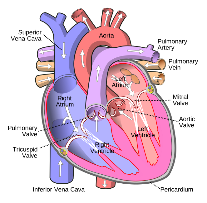
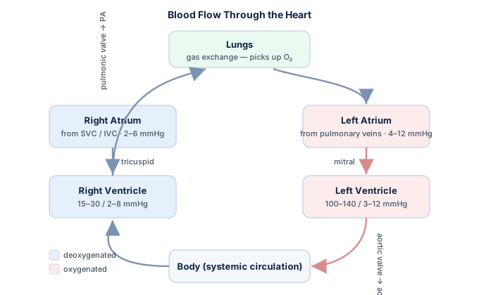
Chambers, valves & blood flow
| Structure | Function |
|---|---|
| Right atrium (RA) | Receives systemic venous blood (SVC, IVC, coronary sinus) |
| Tricuspid valve | RA → RV |
| Right ventricle (RV) | Pumps to the lungs |
| Pulmonic valve | RV → pulmonary artery |
| Left atrium (LA) | Receives oxygenated blood from 4 pulmonary veins |
| Mitral (bicuspid) valve | LA → LV |
| Left ventricle (LV) | Pumps to the body (highest pressure) |
| Aortic valve | LV → aorta |
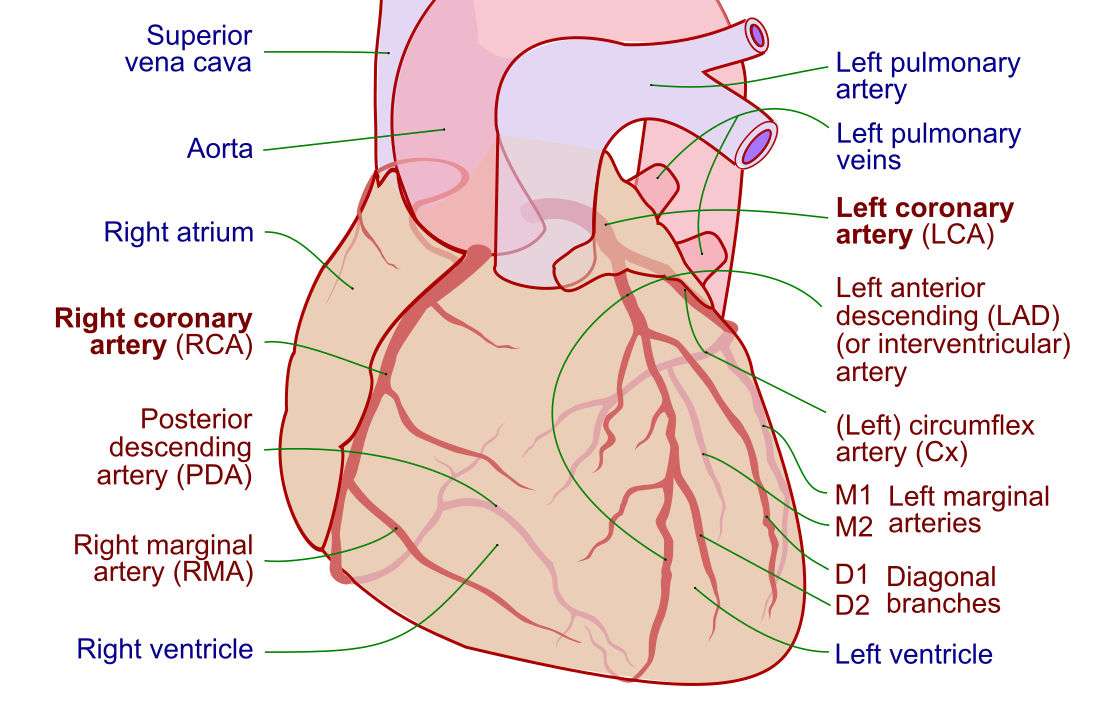
Coronary arteries & territories
| Artery | Supplies |
|---|---|
| Left main (LM) | Divides into the LAD and LCx |
| Left anterior descending (LAD) | Anterior wall, anterior ⅔ of the septum, apex |
| Left circumflex (LCx) | Lateral wall (obtuse marginal branches) |
| Right coronary artery (RCA) | Inferior wall, SA node (~60%), AV node; gives the PDA in right dominance |
The conduction system
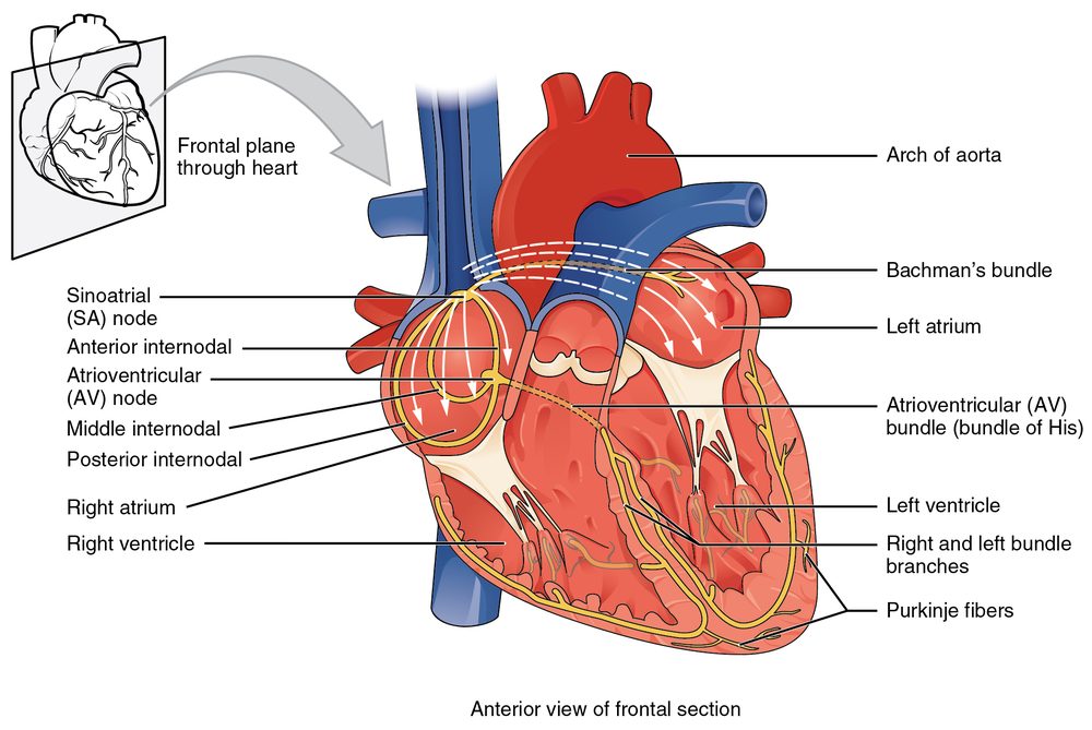
The normal impulse pathway:
| Step | Structure |
|---|---|
| 1 | SA node (pacemaker, ~60–100/min) — high right atrium |
| 2 | Internodal atrial pathways → atrial contraction |
| 3 | AV node — the normal delay allows the atrial kick |
| 4 | Bundle of His — the only normal atrio-ventricular connection |
| 5 | Right & left bundle branches → Purkinje fibers → ventricular contraction |
Study these in depth with the anatomy question bank, the coronary anatomy guide, and the conduction system guide.
Frequently asked questions
Which coronary artery supplies the inferior wall?
In most people the right coronary artery supplies the inferior wall (and the SA and AV nodes), which is why inferior MIs are often RCA occlusions.
What is coronary dominance?
Dominance describes which artery gives rise to the posterior descending artery: right-dominant (RCA) in about 85% of people, left-dominant (circumflex) in about 8%, and co-dominant in about 7%.
What is the normal cardiac conduction pathway?
SA node → atrial pathways → AV node (delay) → bundle of His → right and left bundle branches → Purkinje fibers → ventricles.
Can I print this cardiac anatomy poster?
Yes — use the 'Print / Save as PDF' button at the top.
Sources & further reading
- Cardiovascular Credentialing International (CCI)
- American College of Cardiology
- American Heart Association
- MedlinePlus (U.S. National Library of Medicine)
External links are provided for reference; always confirm current details with the official source.